


-30%


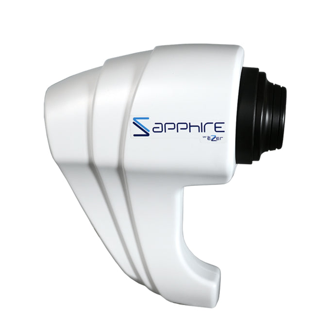
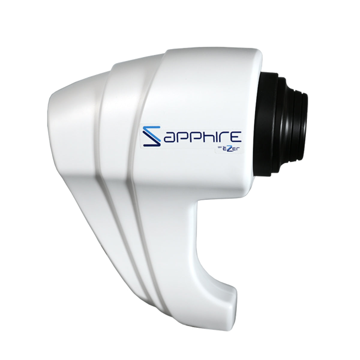
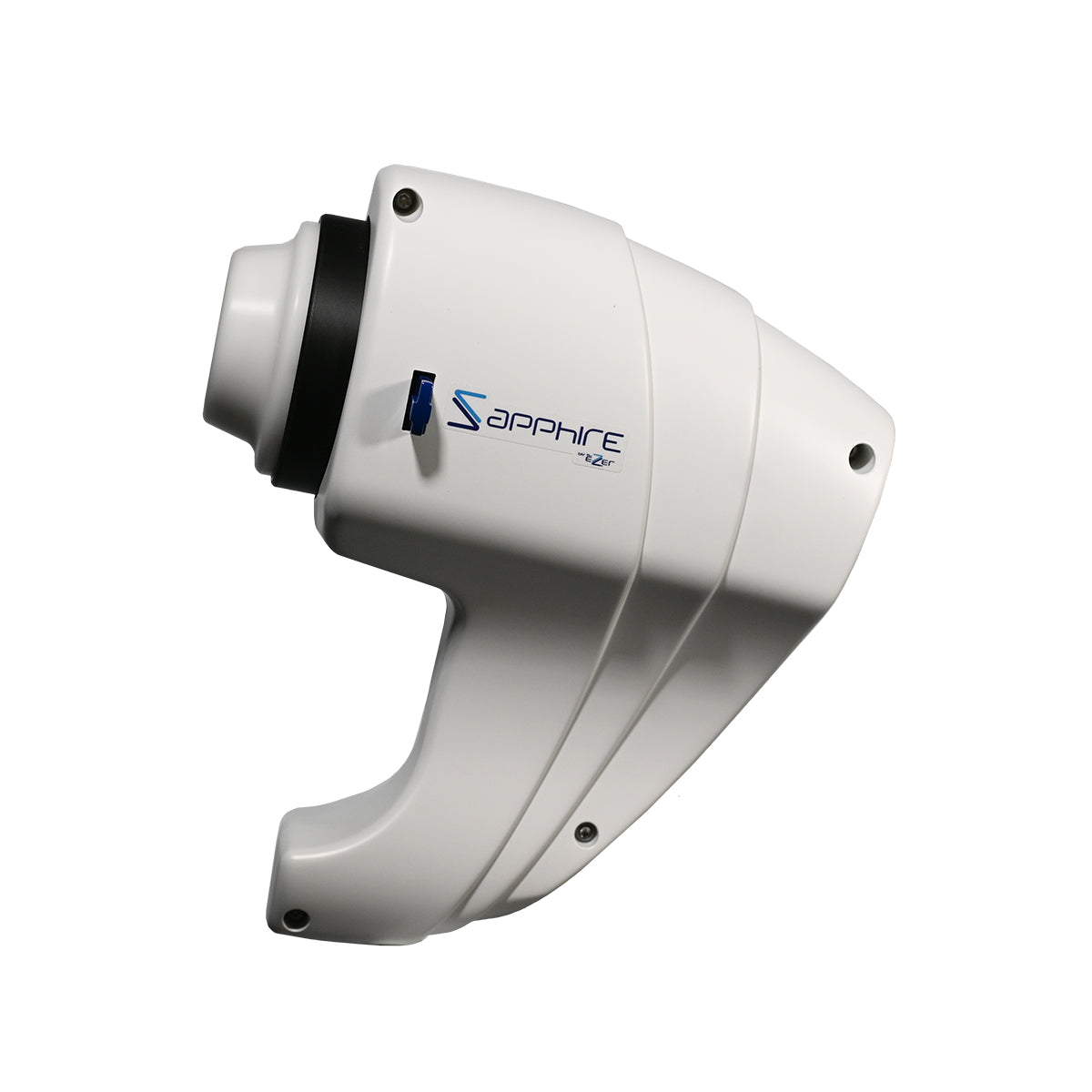
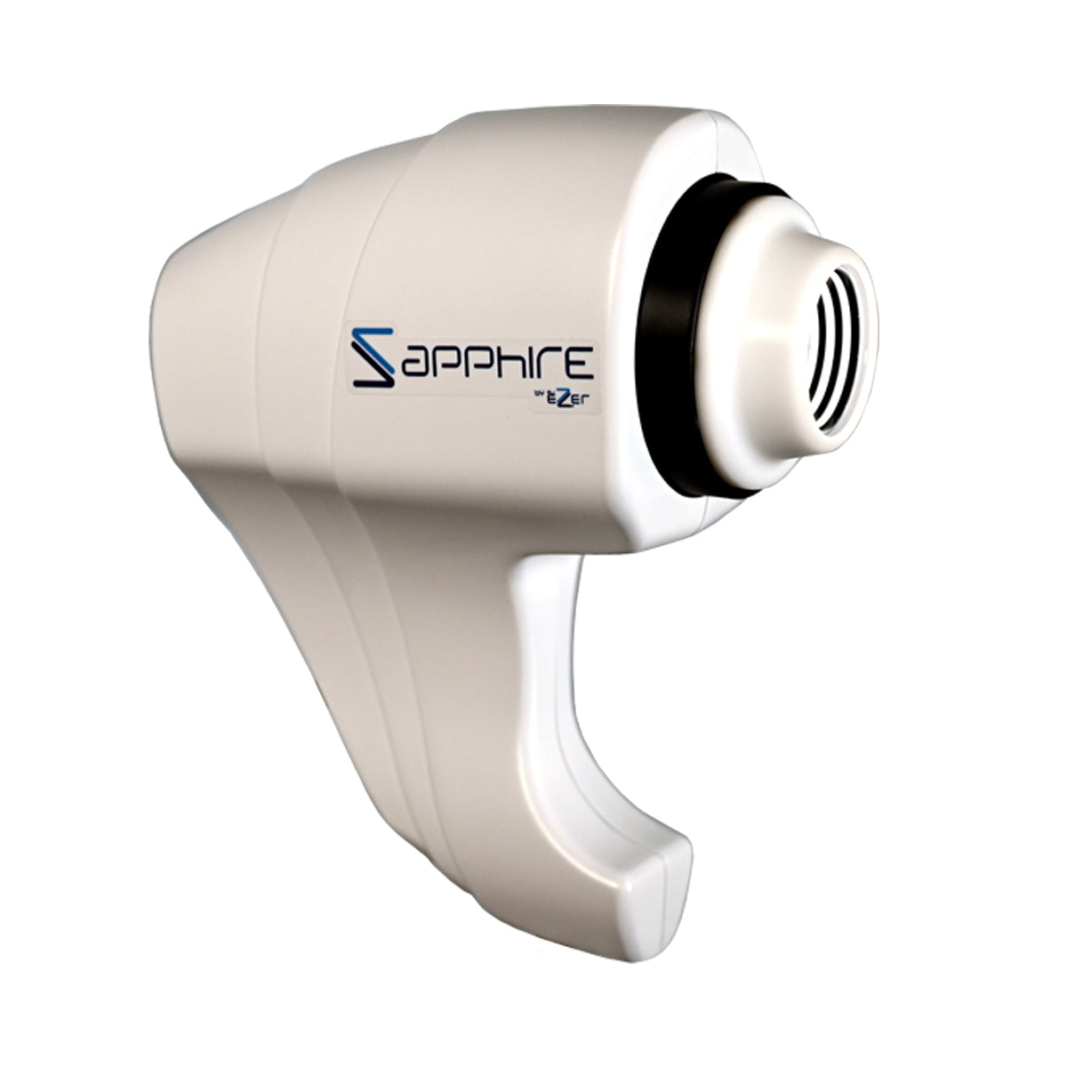
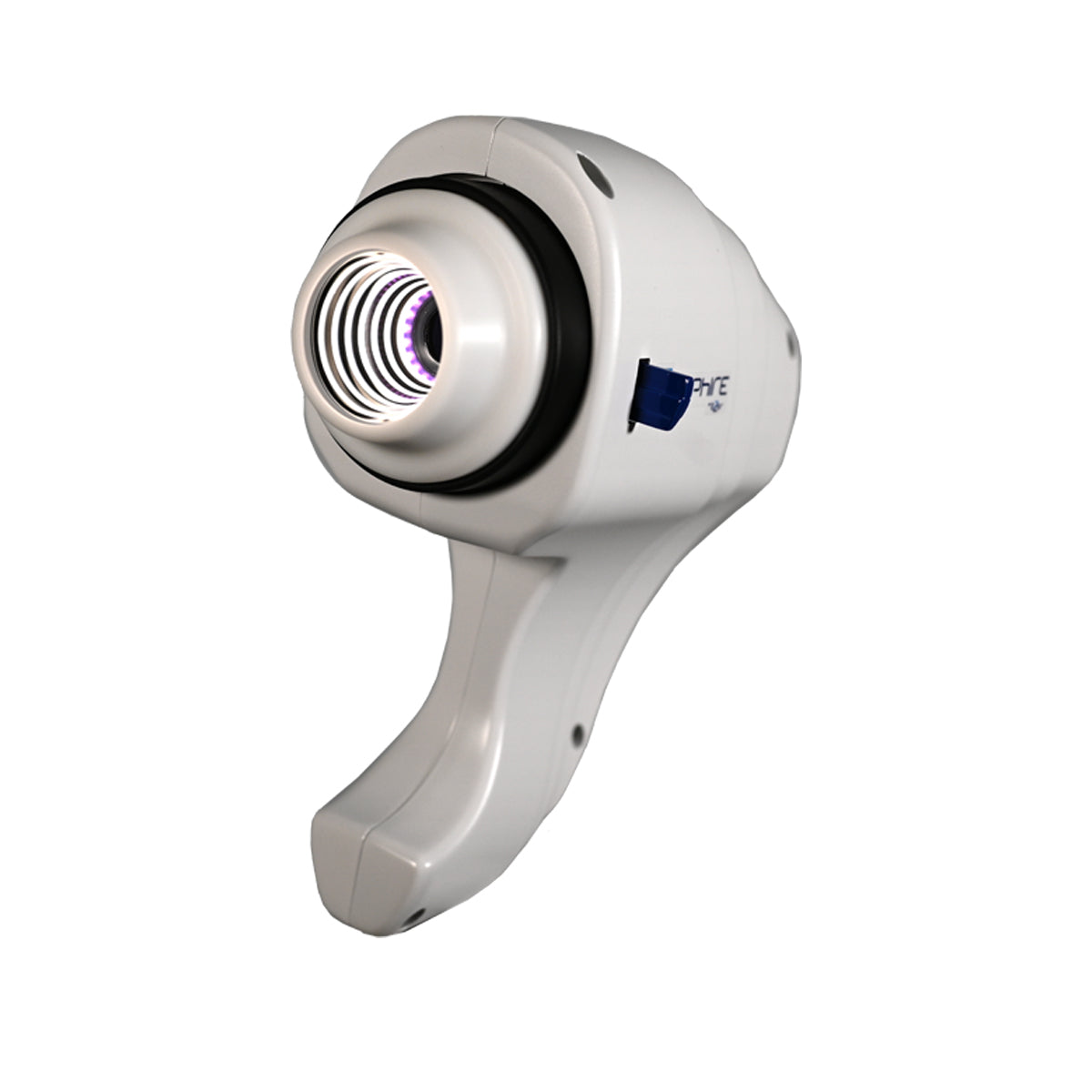
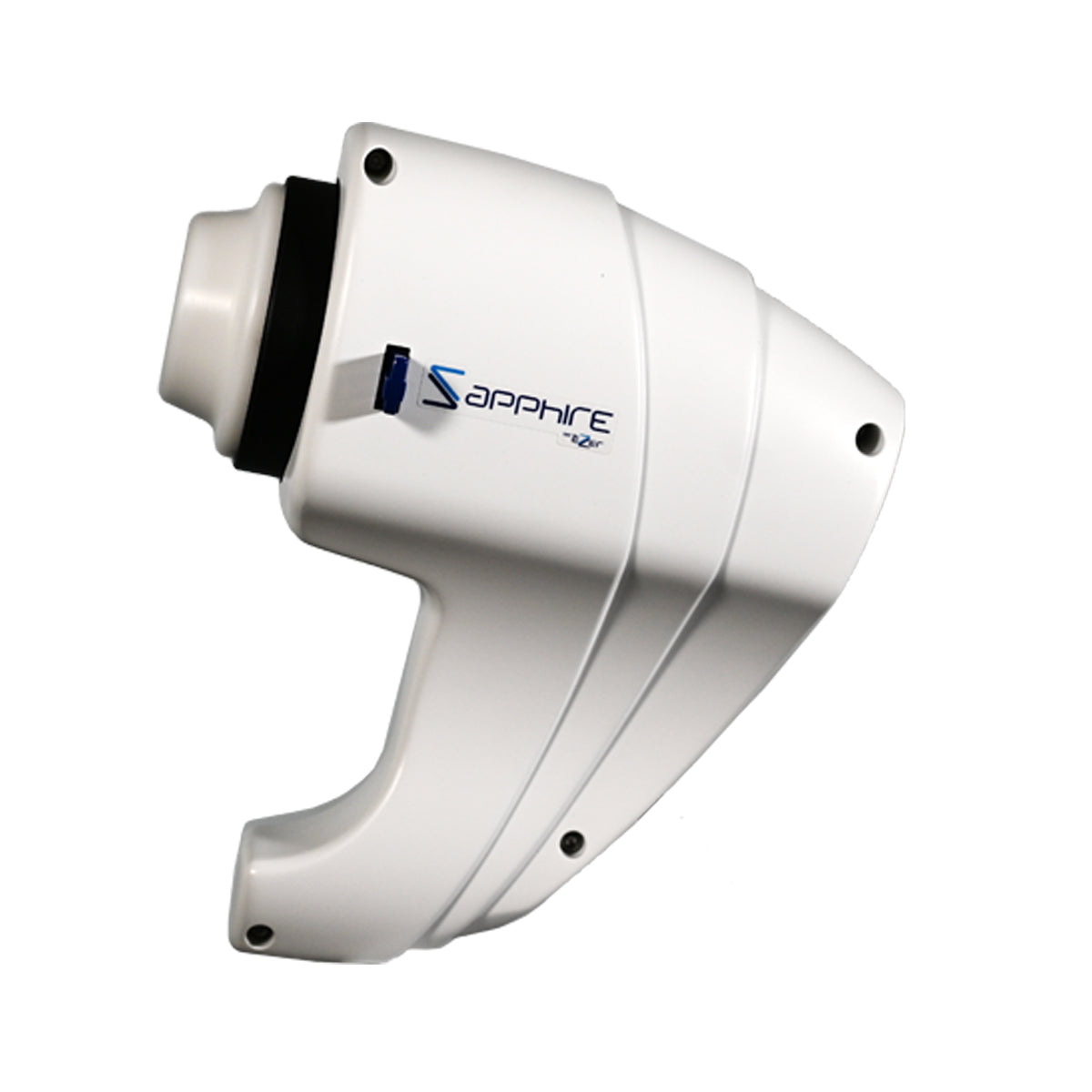

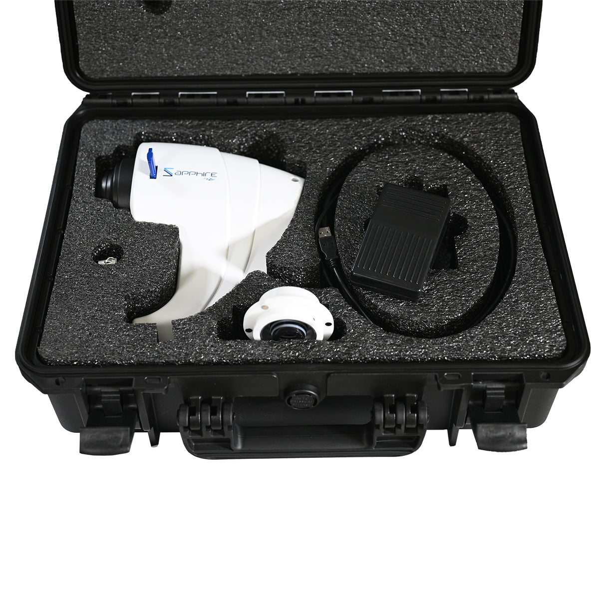
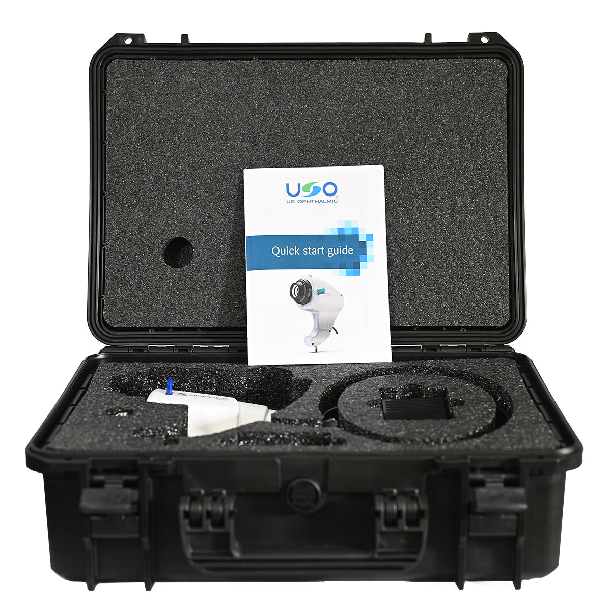
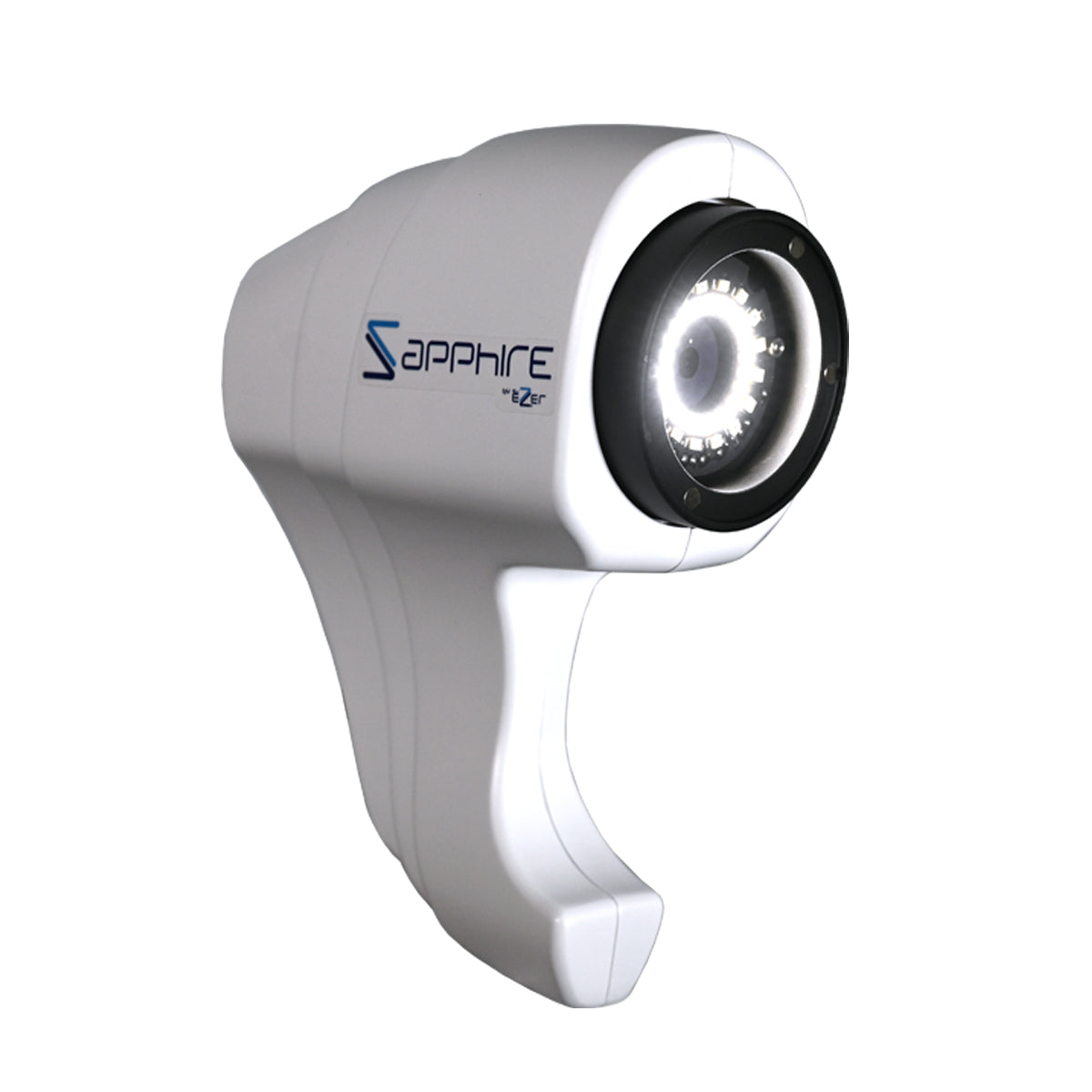
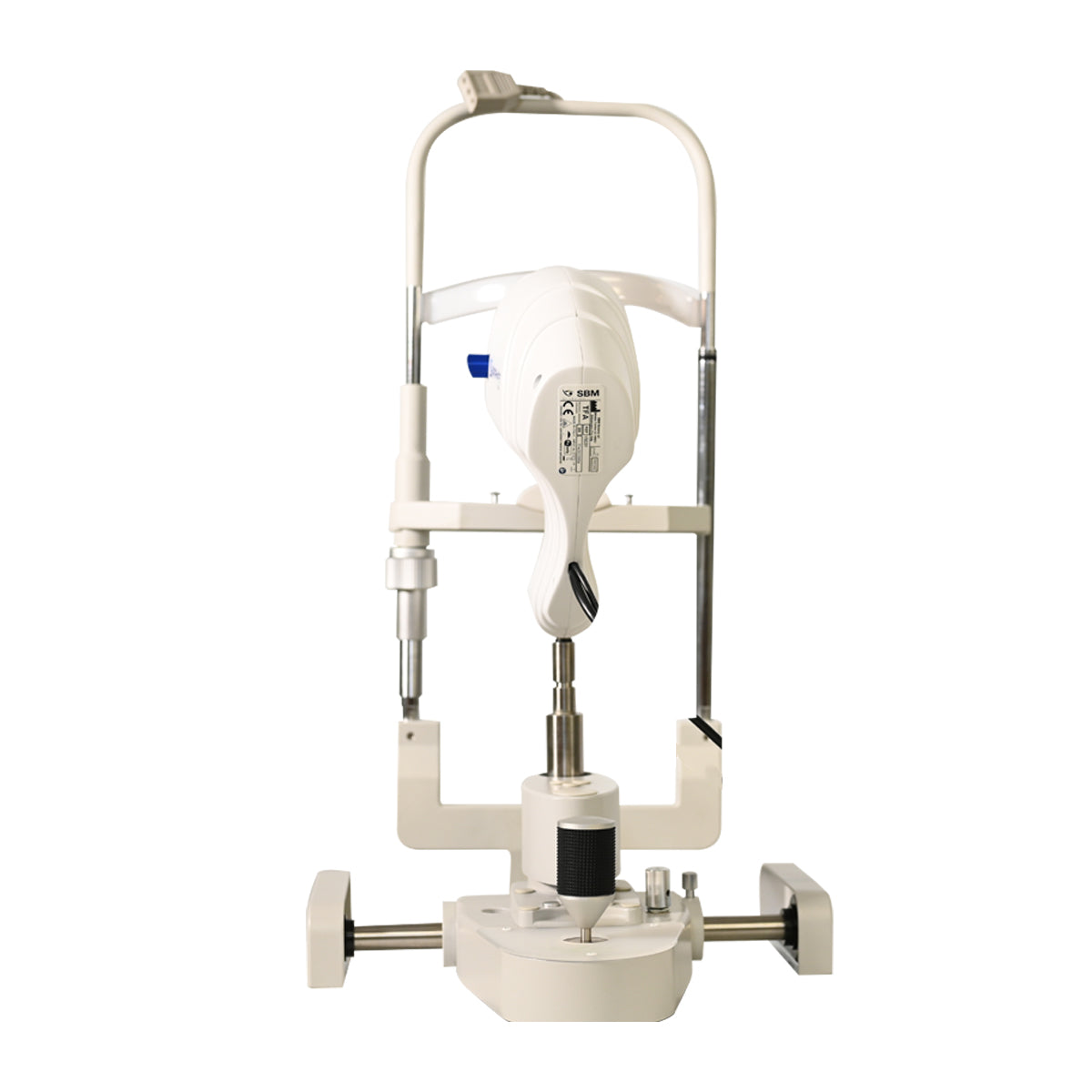
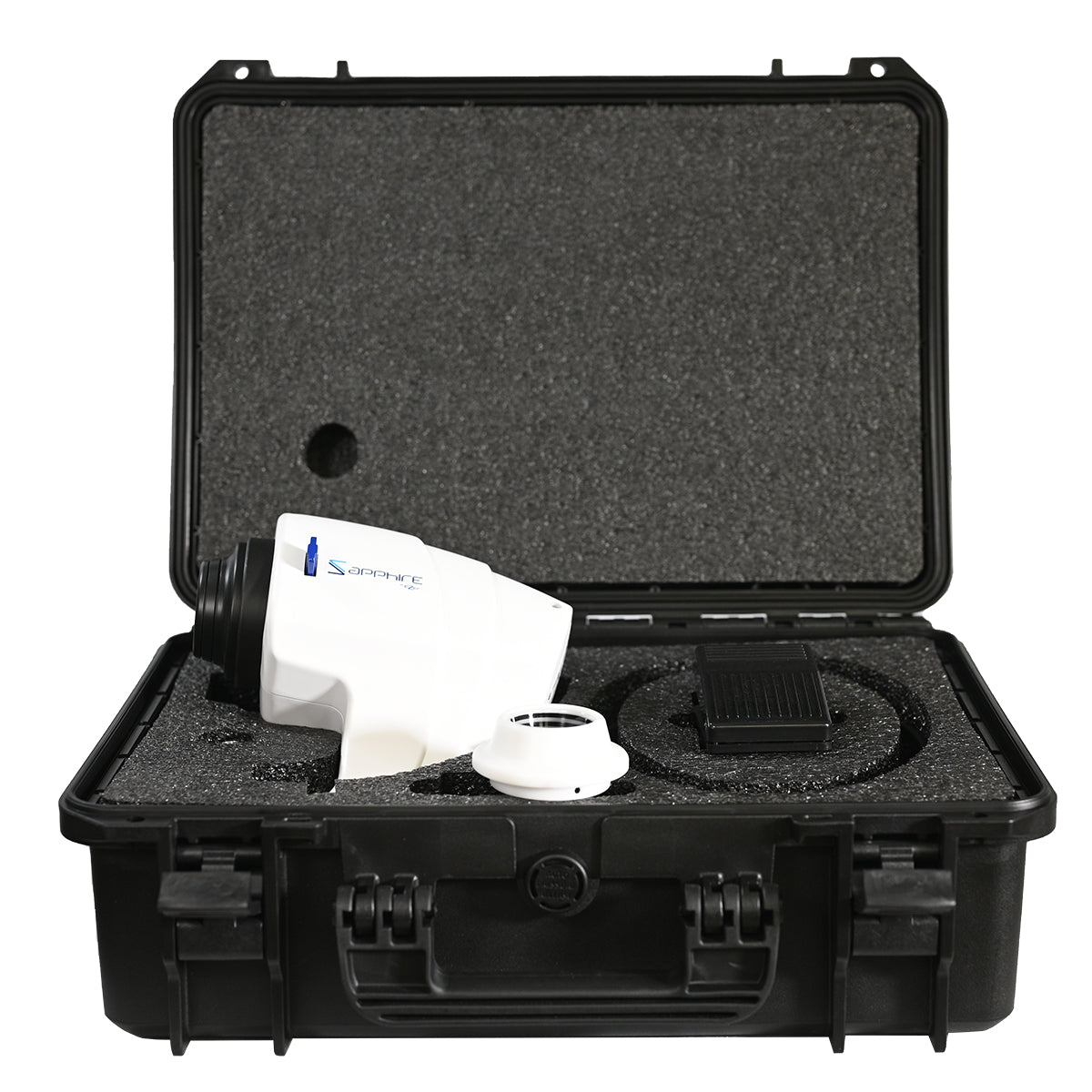
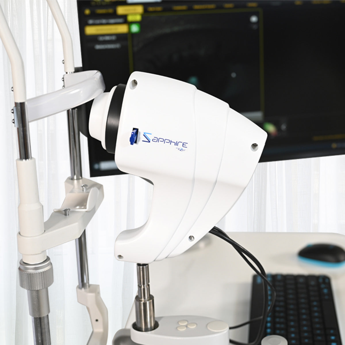
Ezer Dry Eye Sapphire
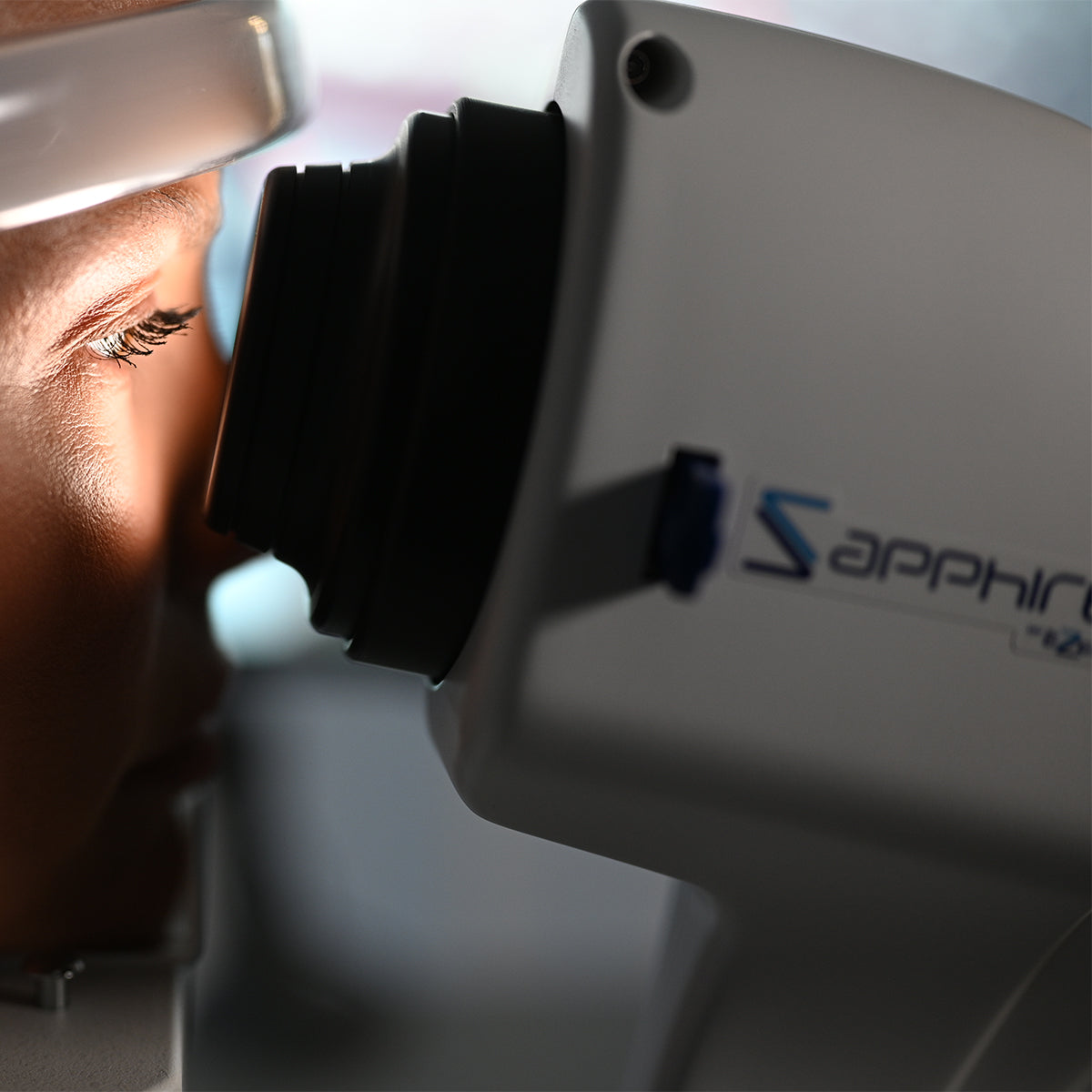
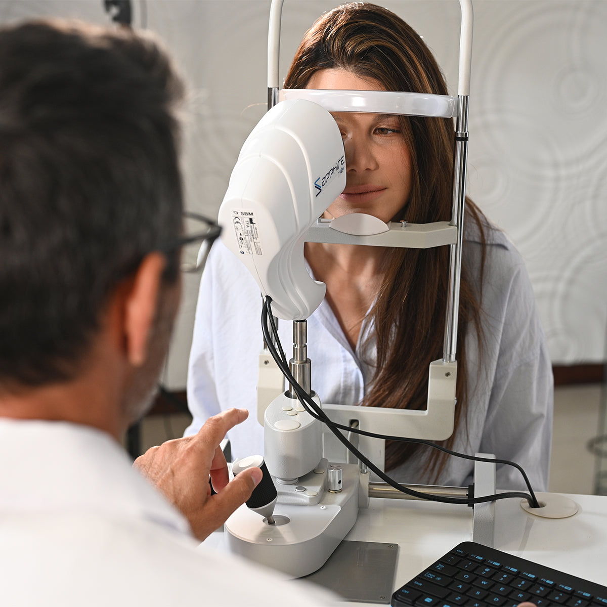
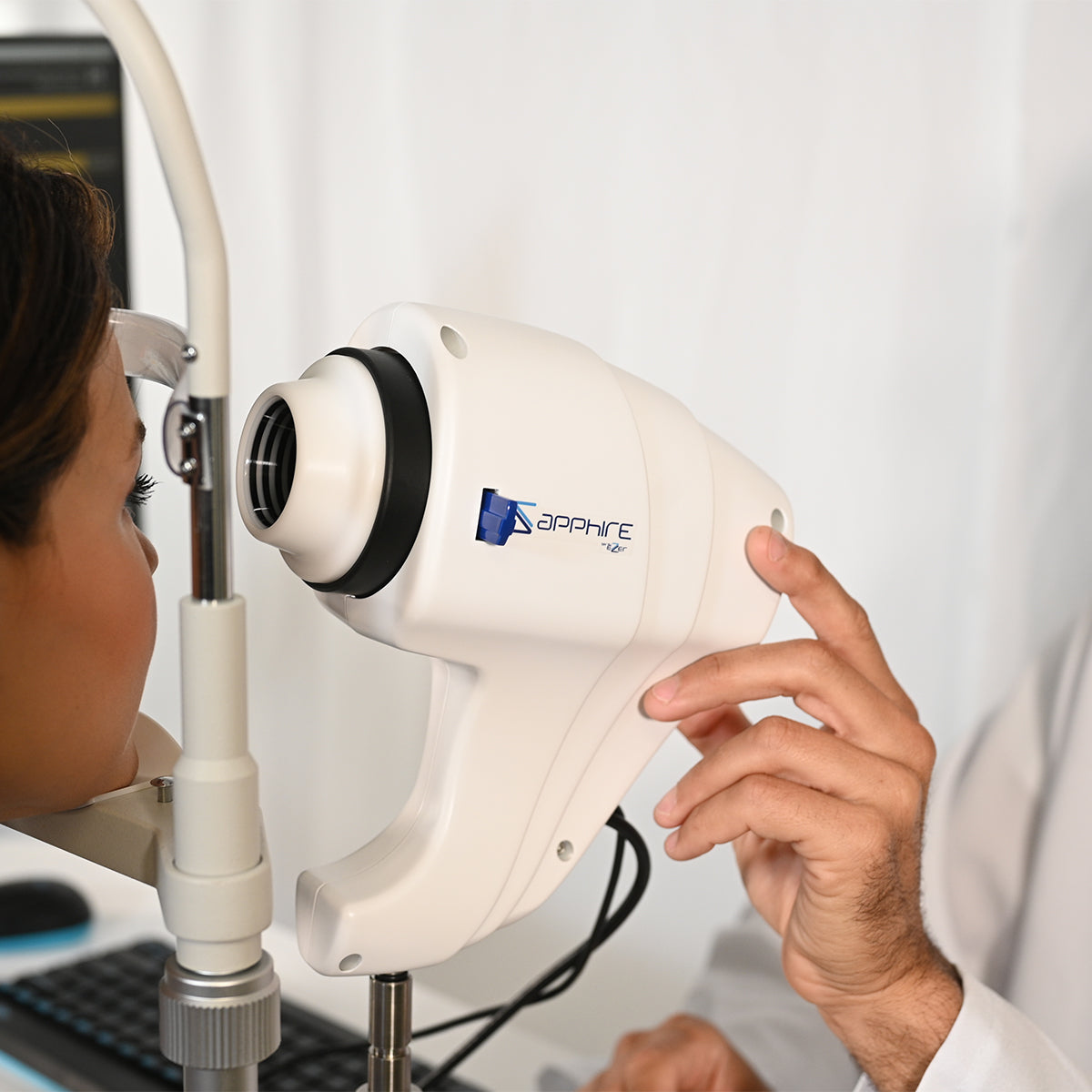
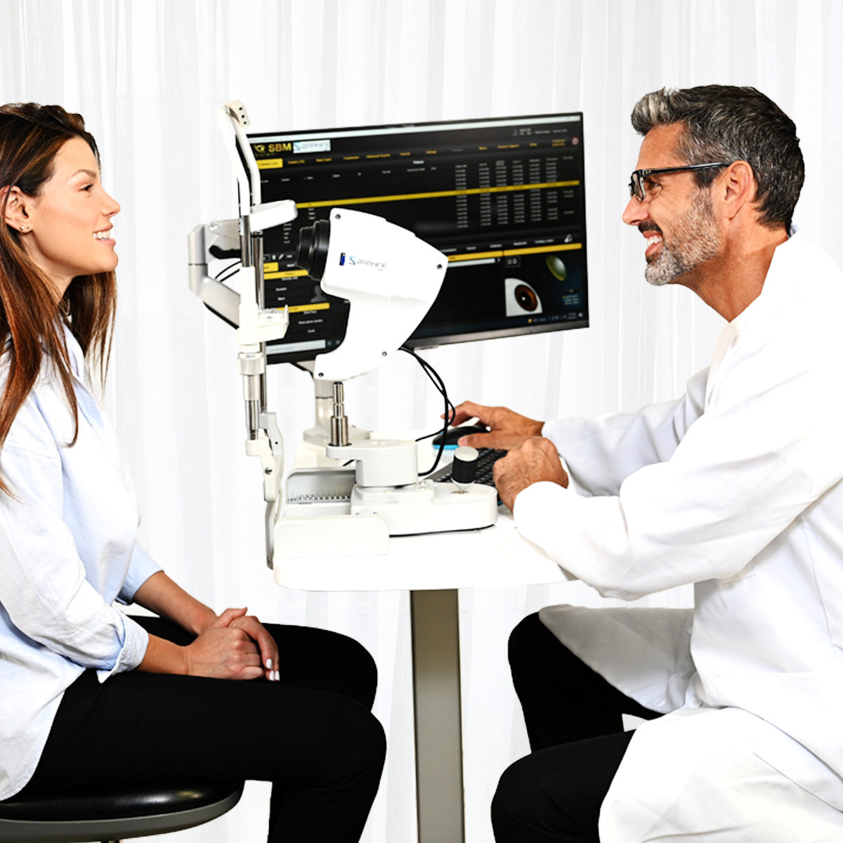
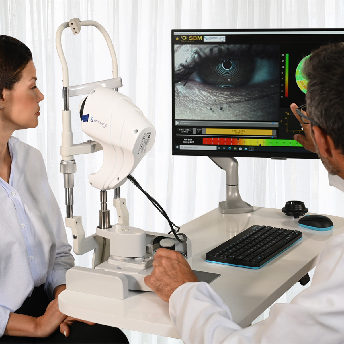
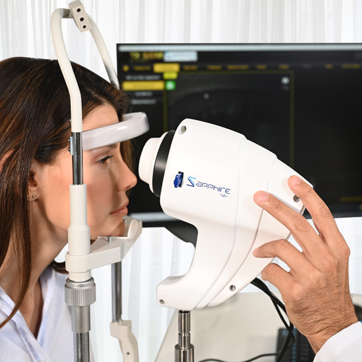
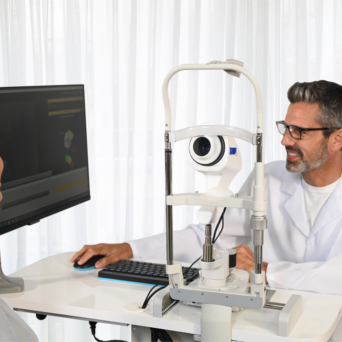
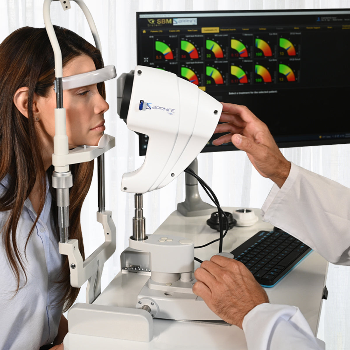
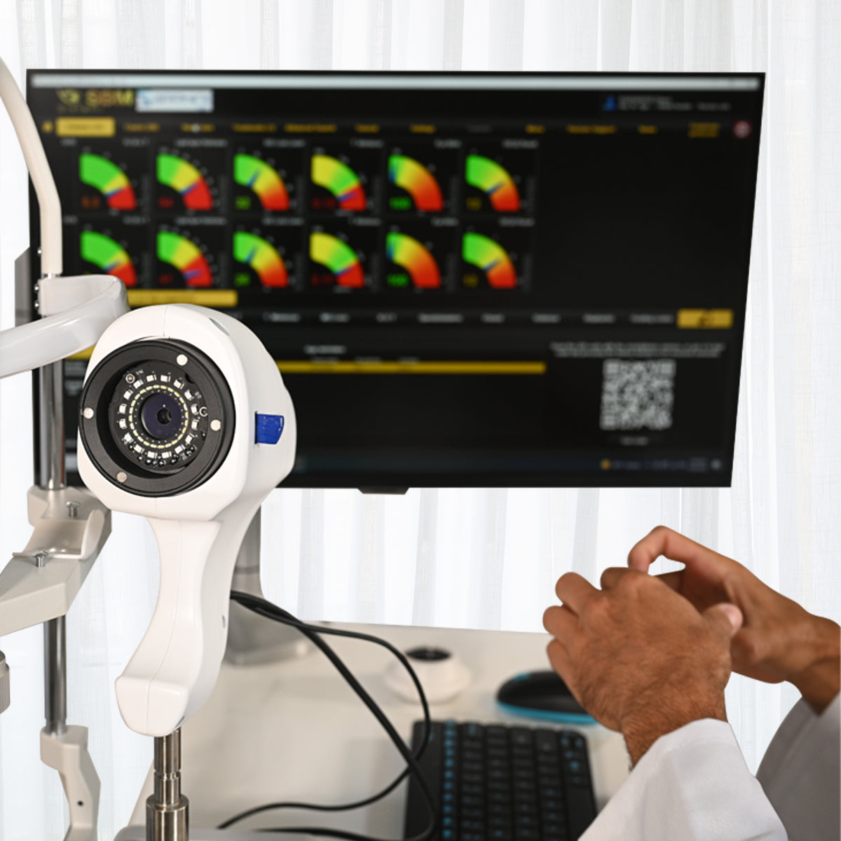
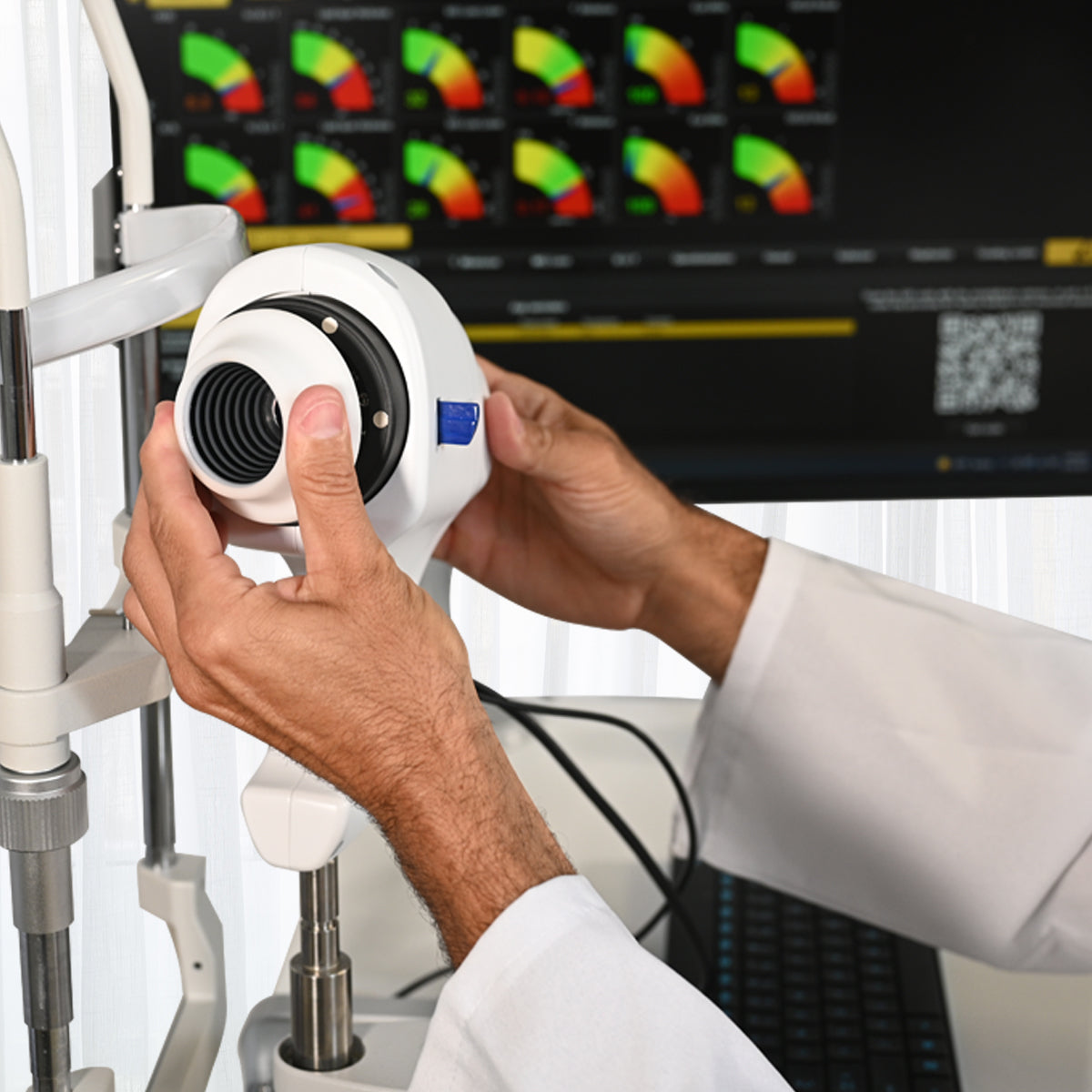
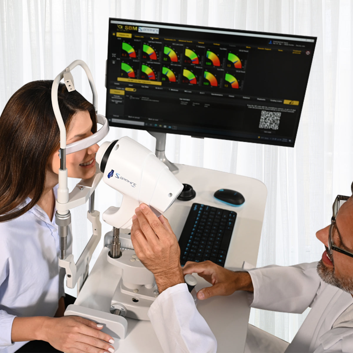
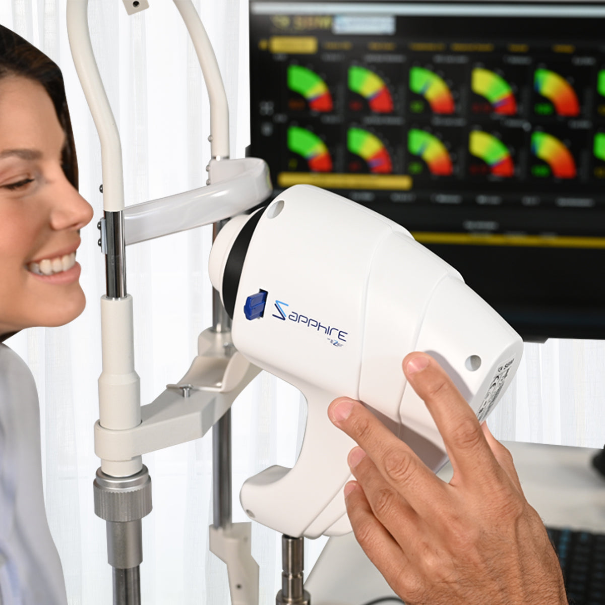
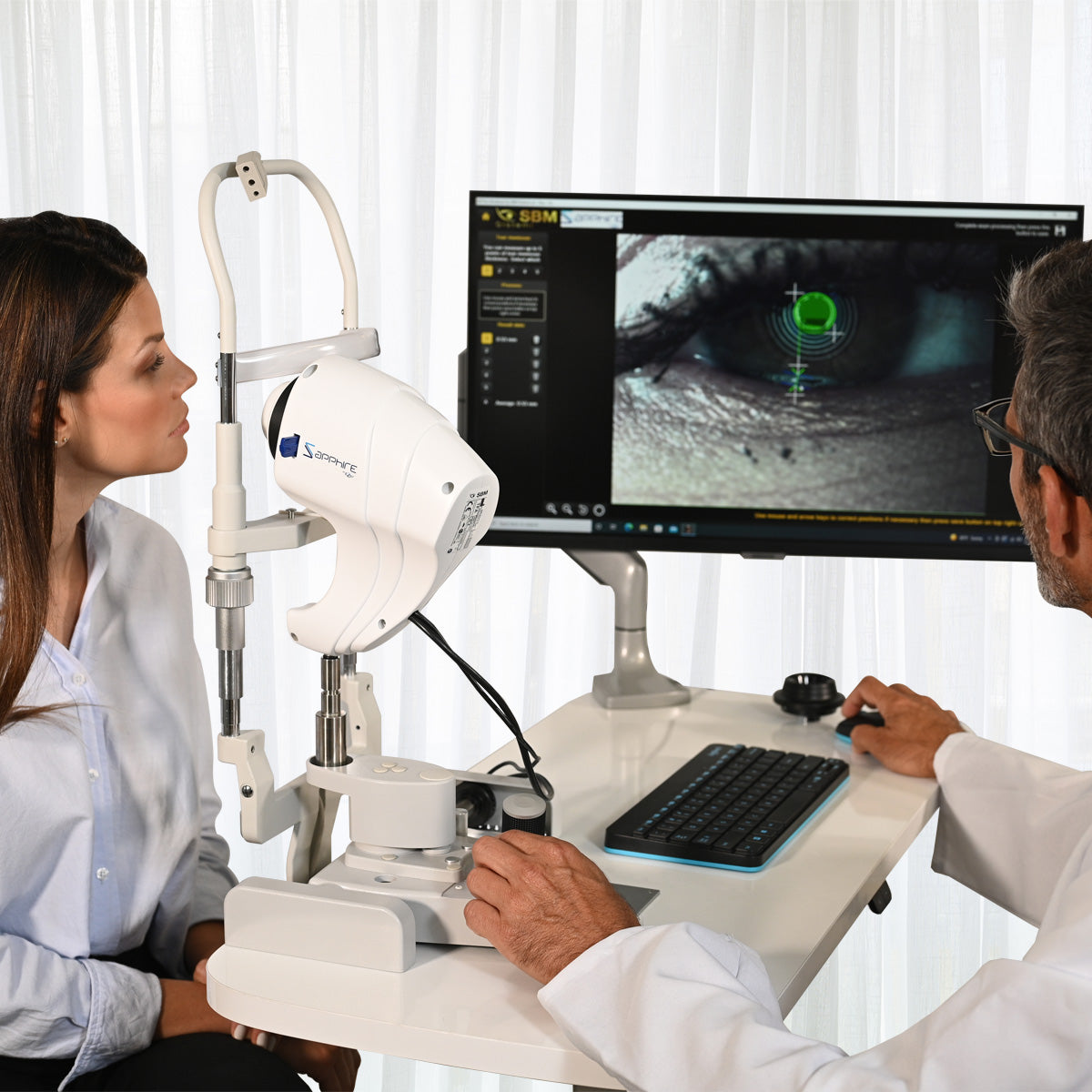
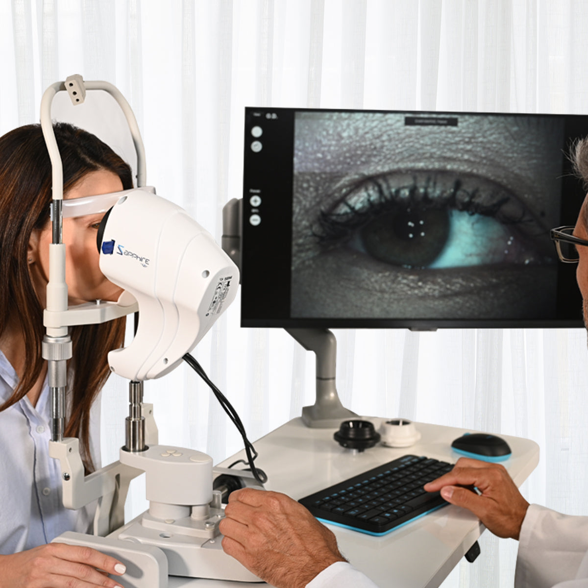
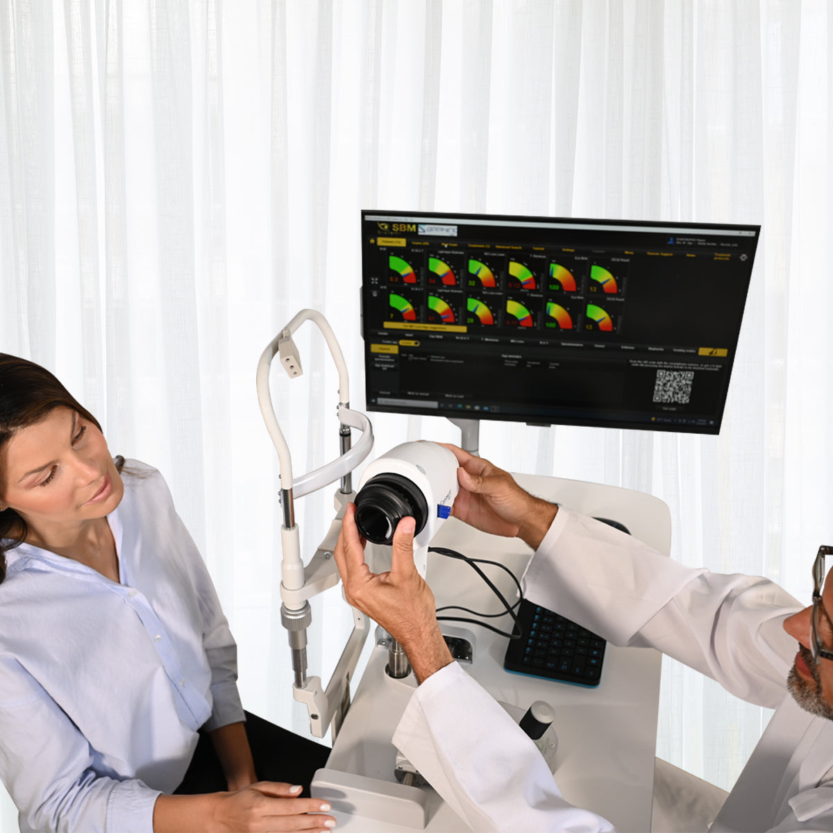
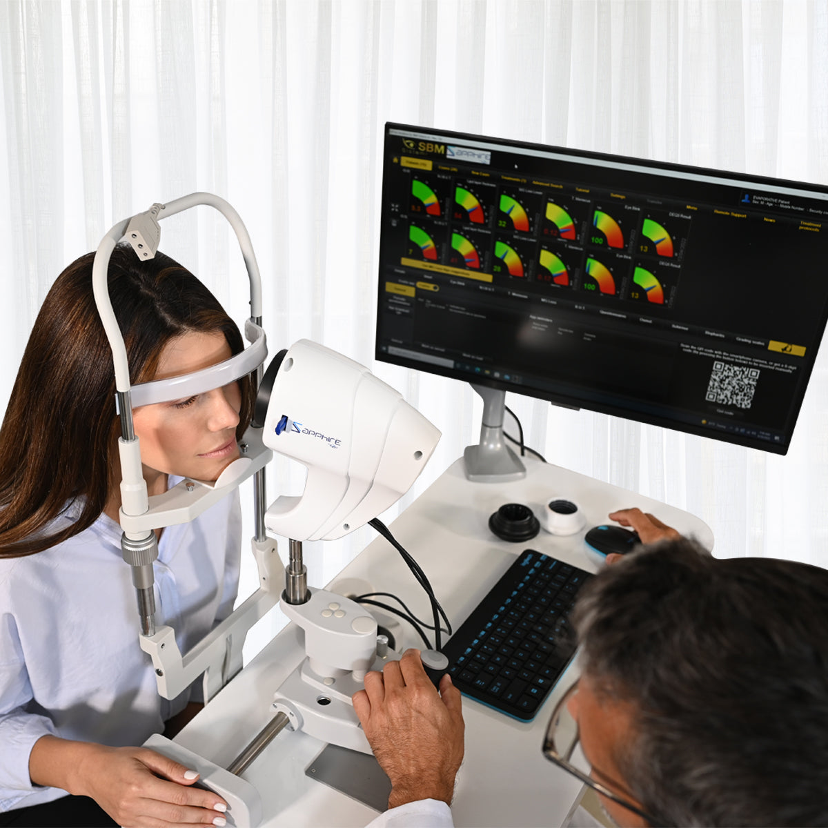
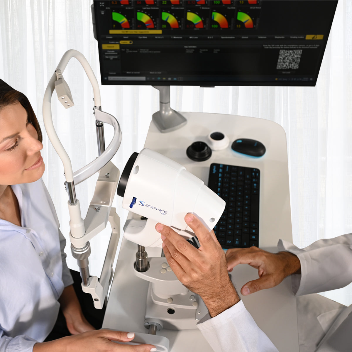
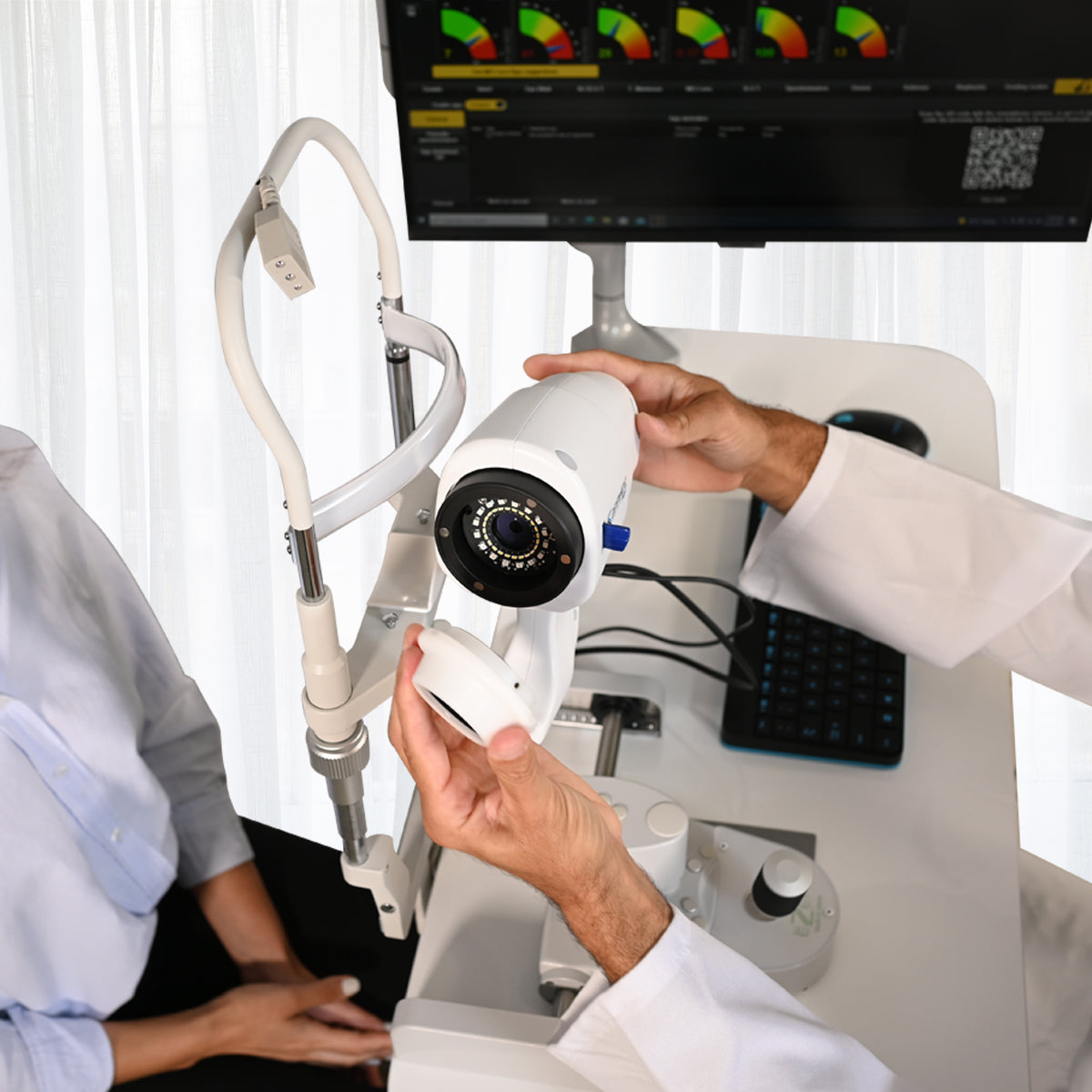
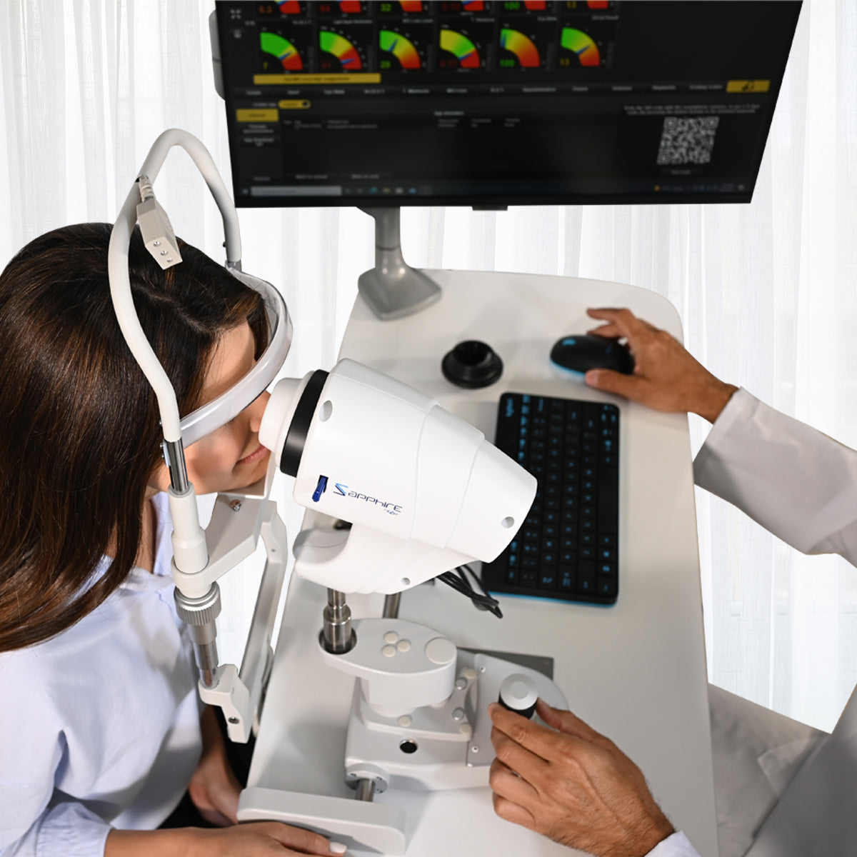
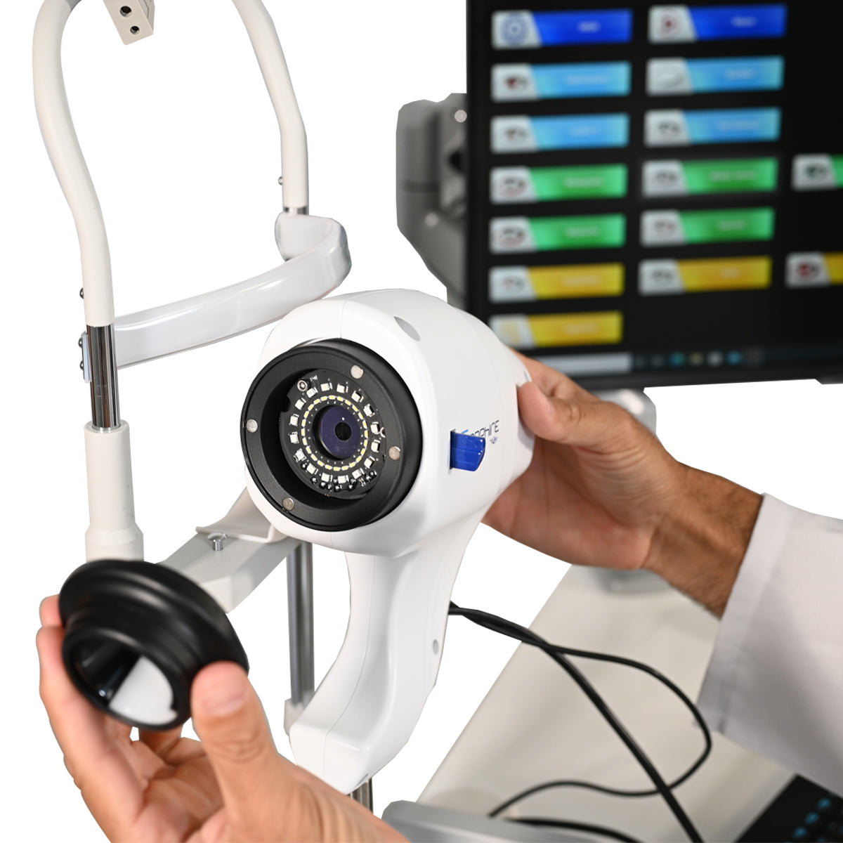
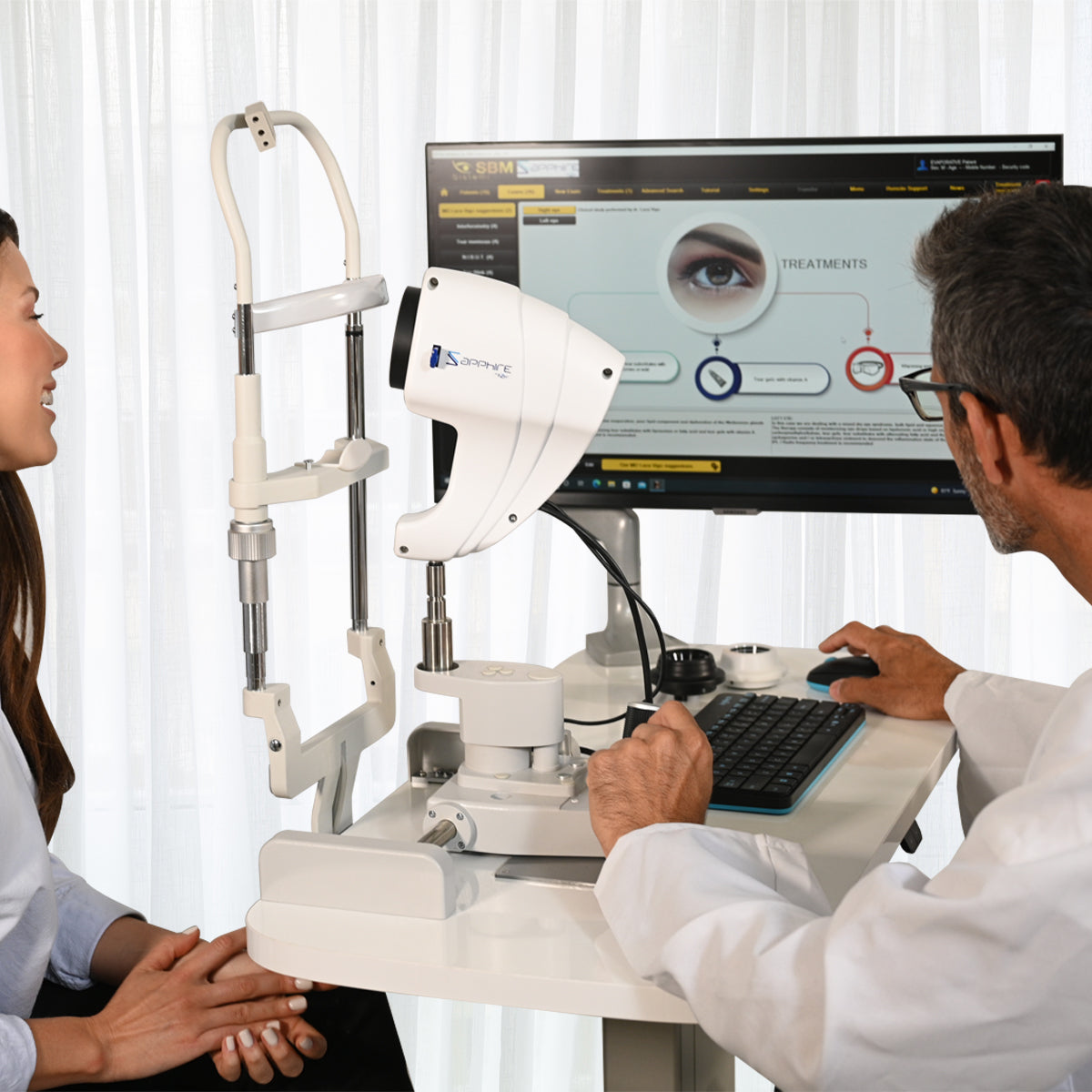
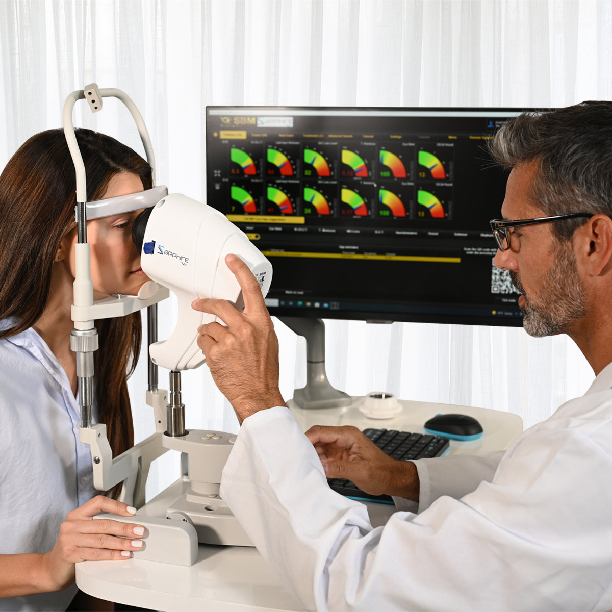
The Sapphire device can therefore provide you with an exceptionally detailed overview of your patient’s tear film composition. It can aid in the diagnosis of the type and severity of the dry eye disease and will recommend a suggested treatment protocol for the individual eye. The device can analyse a number of different features, including the lipid layer, tear meniscus, blood vessels and more. The Sapphire device can therefore provide you with a full picture of your patients’ eye health, so you are able to determine the cause and treatment for dry eyes.
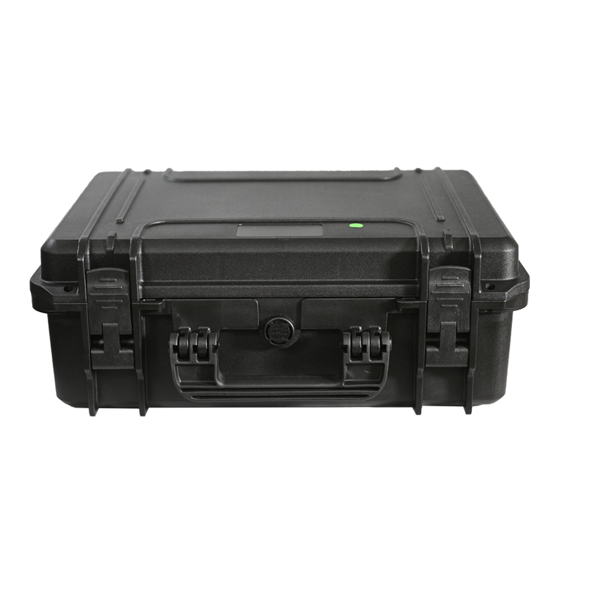
*Purchase includes one software license
The Sapphire is designed to perform tear film tests, from the quality of tears to the analysis of the Meibomian Glands. Is the new instrument for the individual analysis of the tear film that allows to carry out a quick detailed structural research of the tear composition.
All in One

Evaluation of all Tear Film Layers

Fully Automatic Protocol

INTERFEROMETRY

Its pin has been built in order to fit perfectly into the hole that you can see when the plate used for the tonometer is removed.
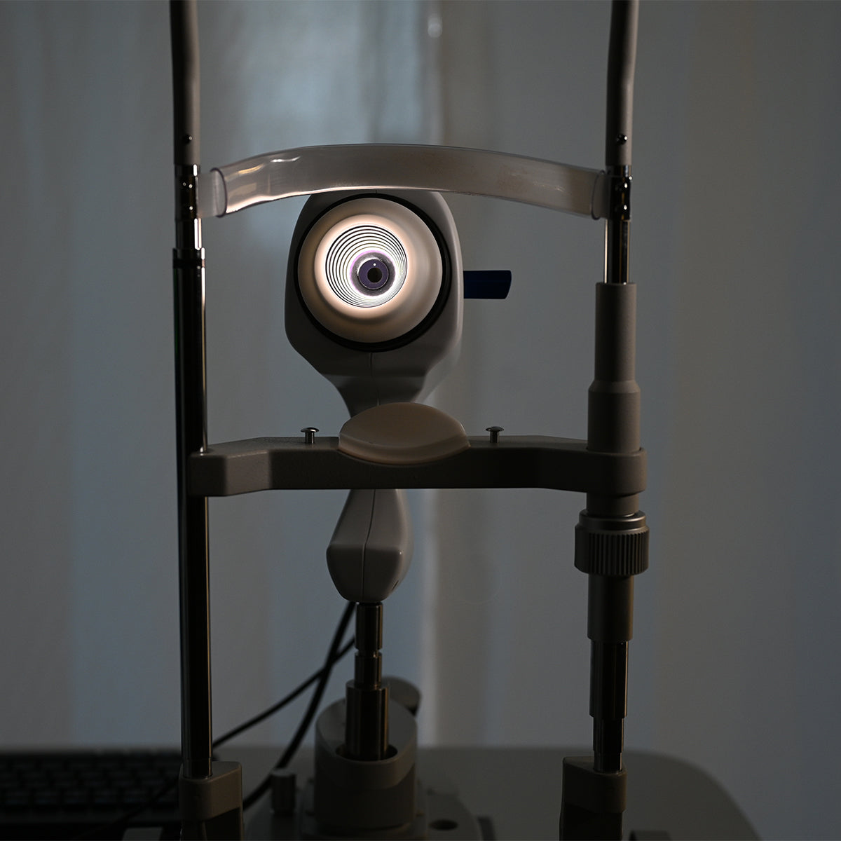
Closer the device is to the eye broader the area is illuminated by the instrument.
The light reflected from the tear film can be observed as a white circular area that almost completely covers the cornea.

This exam has fundamental importance, because most of the dry eye diseases are caused by an insufficiency of lipid layer quantity.
The evaluation of the lipid layer is part of your overall Dry Eye Assessment. Knowing what is causing Dry Eye will help determine the best treatment option.

LIPID LAYER ANALYSIS
LLT measurements are presented in an easy to understand color-coded scale. The identification is done through the international grading scale of Dr. Guillon.
TEAR MENISCUS
Low tear production may result in aqueous tear deficiency (ATD) and cause dry eye symptoms.
However, measuring the tear volume is difficult since the methods available nowadays are invasive and irritating.
NIBUT
Thanks to one single video the physician can gain lots of information:
• Automatic NIBUT
• Average of more than one value
• Graph to understand the trend of tear film stability during the video
• Tear topography that shows all breaking the tear film during time.
MEIBOGRAPHY
Meibomian Glands (MGs) play a significant role in tear quality by producing lipids (meibum) that are part of the superficial tear film. Dysfunction of the MGs destabilizes tear composition resulting in evaporative dry eye.

The System automatically analyses the images taken through a sensitive infrared camera (NIR) to locate the Meibomian Glands in a guided way:
• An exam valid both for the upper and the lower eyelids;
• Automatic percentage of the Meibomian Gland loss area

Meibomian Gland dysfunction (MGD) is characterised by chronic, diffuse abnormalities of the Meibomian Glands and altered secretion and chemical composition of meibum.
BLEPHARITIS AND CYLINDRICAL DANDRUFF

CYLINDRICAL DANDRUFF AND BLEPHARITIS The human skin surface is known to house millions of bacteria, though some people have more than the average number. Blepharitis is an inflammation caused by some bacteria that lie at the base of eyelashes. They produce dandruff-like flakes in the skin, which lead to infection and inflammation.

Problems with the Meibomian Glands (meibomianitis) in the eyelids can also cause blepharitis. The development of inflammation is also associated with risk factors such as dandruff, dry eye, acne rosacea, or bacteria.

This test helps in the detection of blepharitis. It can be performed on the outer surface of the eyeball and eyelids.
The process includes:
• Examining the openings of the Meibomian Glands, base of the eyelashes, and eyelid margins using a bright light.
• Checking for abnormalities by evaluating the quantity and quality of tears.

The type of blepharitis can be determined based on the appearance of the eyelid edges. If the symptoms frequently exhibited by the patients are mildly sticking eyelids, thickened lid margins, and missing/ misdirected eyelashes, then the type of blepharitis is diagnosed as Staphylococcal.

When the patient is found with blockage of the Meibomian Glands in the eyelids, poor quality of tears, and redness of the lining of the eyelids, Meibomian blepharitis is diagnosed.
If a hard, matted crust is formed on the eyelashes, and after its removal small sores appear on the skin, Ulcerative blepharitis is diagnosed.
WHAT IS DEMODEX BREVIS?
Demodex brevis is a kind of mite found on the skin of humans.
Like its counterpart Demodex folliculorum, D. brevis is naturally occurring. D. brevis is so small that mitas can’t be seen macroscopically.
MD LUCA VIGO SUGGESTIONS
Suggestions for diagnosis and treatment based on clinical procedure by Dr. Luca Vigo and studio carones (Milan, Italy).

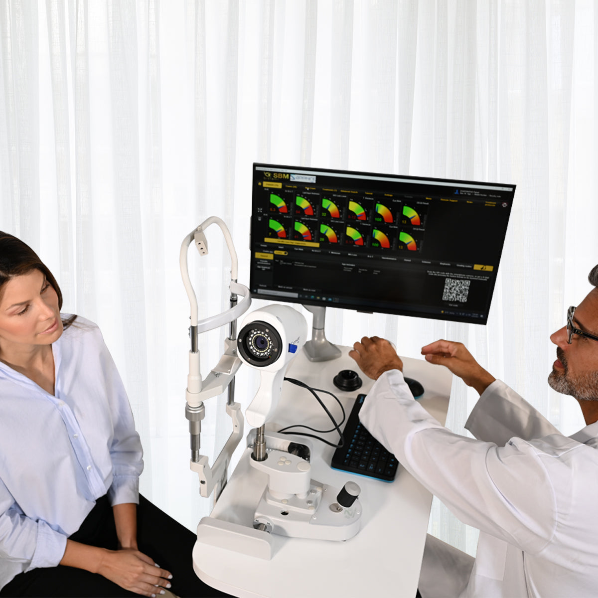
A complete and dry eye-focused database allows to understand and properly diagnose the dry eye patient. With the useful data result tab, the ophthalmologist can check the complete tear film assessment, determining all deficiencies that cause the pathology and meantime understanding which treatment is needed to approach each case.

DIAGNOSIS SUGGESTION
Ocular surface data and pathology classification
Thanks to Studio Medico Carones with MD. Luca Vigo’s experience, TFA includes a suggestion algorithm able to share a possible treatment approach for each patient.

TREATMENT MANAGING
Through TREATMENT MANAGING tab, the software allows the physician to fill in the database with all drugs, integrators and treatmenrs available in his practice.
Any treatments as vitamins, Omega-3, eye drops, hot packs and IPL/Radiofrequency, can be loaded on the software to prescribe the products of the brands that the doctor prefers.
The whole report with the diagnosis and treatment suggested by the ophthalmologist will be printed directly.

PROTOCOL
(MD L.VIGO Studio Carones Milano) is already provided and suggest treatment on our KOL experience.

All users can customize their own protocol adding treatment procedure to be chosen automatically right after performing the exams (this makes it possible as well to delegare the diagnosis to an assistant).
OTHER POSSIBLE EXAMINATIONS

AN ASSESSMENT OF GRADING SCALES FOR MEIBOGRAPHY IMAGES
The evaluation of the Meibomian Gland dysfunction appears to be of increasing interest in research and clinical practice. Consequently, the evaluation of MGs morphology using Meibography is of high interest for both researchers and clinicians.

WHITE TO WHITE MEASUREMENT
Evaluation of corneal diameter from limbus to limbus (white-to-white distance, WTW).
BULBAR REDNESS CLASSIFICATION
Acquiring an image of the conjunctiva, it will be possible to compare the patient’s condition with different international grading scales.


PUPILLOMETRY
The measurement of the pupil diameter has become increasingly important in the field of refractive surgery.
Larger scotopic pupil sizes may be partially responsible for the occurrence of postoperative symptoms such as halos, glare, and monocular diplopia.
Refractive surgeons also need an accurate scotopic pupil measurement to determine appropriate treatment zones for excimer laser, corneal, and intraocular surgery.

COMPARISON WITH THE MAIN INTERNATIONAL GRADING SCALES
EFRON - CCLRU - JENVIS - GLAUCOMA - FERNING TEST - MEIBOGRAPHY
DIFFERENT REPORTS AVAILABLE
The TFA software is a dedicated platform for dry eye and allows, in addition to helping in the diagnosis and classification of diseases, to print and save various medical reports, offering the most professional and clinical solutions to patients.
For customer satisfaction, it is often advisable to provide technical documentation relating to the exams taken.
Thanks to the various print reports of the Sapphire device, you will have the possibility to visually explain and simply demonstrate the pathology situation. Furthermore, it’s possible to explain how the pathology has changed over time.

COMPLETE REPORT
Complete report with all results and pictures used to explain to the patient any dry eye category.

TREATMENT REPORT
Patient oriented report explaining causes of pathology and recommended treatments.

FOLLOW UP REPORT
For each value it is possible to show the trend line before/during/after treatment.

DAILY REPORT
Brief single page report to show at a glance all exams results.

MONOCULAR REPORT
To save and prind one only interesting examination.

BINOCULAR REPORT
To save in a single pdf the same eamination of both eyes.
The Sapphire device provides 3D imaging, to help you explain the cause of dry eye and assist your recommendations for treatment. It gives you the ideal tool to easily explain what the cause of the patients’ eye discomfort is, while helping each patient to understand where your recommendation is coming from.
The Sapphire device means you can offer peace of mind to your patients, with the knowledge that you are using the latest technology for correct diagnosis and treatment.
Video Sapphire
Sapphire - Dry Eye Disease
| Image Resolution | 5 MP |
| Acquisition Mode | Multi shot, video |
| Focus | Autofocus, manual focus |
| ISO Management | Variable |
| Cones | Main cone and Placid cone |
| Camera |
Colored, sensitive to infrared
(NIR), yellow-filtered |
| Light Source | Infrared LED – Blue, red and white LED |
| Image Resolution | 5 MP |
| Acquisition Mode | Multi shot, video |
| Focus | Autofocus, manual focus |
| ISO Management | Variable |
| Cones | Main cone and Placid cone |
| Camera |
Colored, sensitive to infrared
(NIR), yellow-filtered |
| Light Source | Infrared LED – Blue, red and white LED |
Sapphire Catalog 2022
Dry Eye Disease - Essential Information
Dry Eye - The impact of Dry-Eye on cataract and refraction surgery
Sapphire - Fully Automatic Dry Eye Diagnostic Platform
Sapphire - Patient Information
Sapphire - Reports
Article 1 - A review of meibography for a refractive surgeon
Article 2 - Ocular Surface Workup With Automated Noninvasive Measurements for the Diagnosis of Meibomian Gland Dysfunction
Article 3 - Noninvasive ocular surface analyzer as an adjunct in diagnosis and estimating prevalence of meibomian gland dysfunction: Hospital‑based comparative study
Article 4 - Algorithmic approach to diagnosis and management of post-refractive surgery dry eye disease
Article 5 - Real-World Diagnostic Performance of a Novel Noninvasive Work-up in the Setting of Dry Eye Disease Journal
Article 6 - Qualitative assessment of the tear film in young adults
Article 7 - Analysis of two latest generation in-office therapies for the treatment of dry eye
Dr. Sergio M. Solarino

Dr. Francesco Carones

Dr. Massimo Gualdi

Dr. Luca Gualdi

Dr. Eros Radrizzani

Dry Eye Disease - Sapphire
Establishing a Treatment Protocol - Sapphire
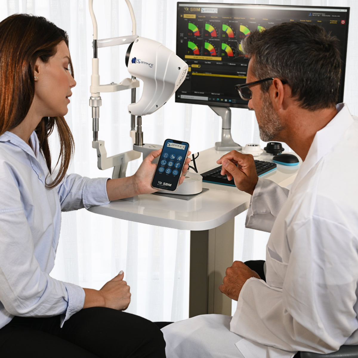
Patient Smartphone Application - Sapphire
Overview of Examination - Sapphire
Understanding NIBUT - Sapphire
Blink Quality - Sapphire
Back to Top

