Retinal and Fundus Cameras
A retinal camera, also called a fundus camera, photographs the back of the eye (the retina, optic disc, macula and vessels) so findings can be documented, compared over time and shared with colleagues. Non-mydriatic cameras capture images through an undilated pupil, which keeps imaging quick enough for routine screening.
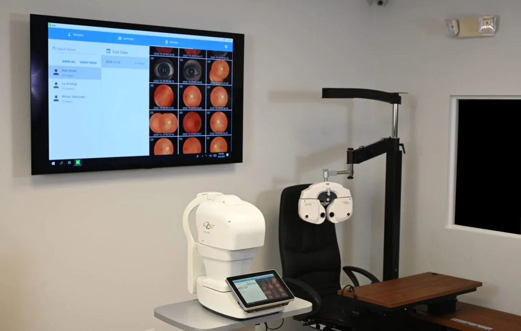
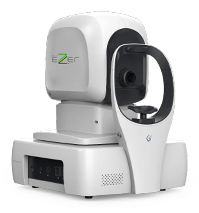
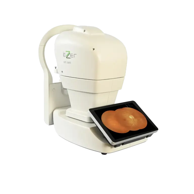
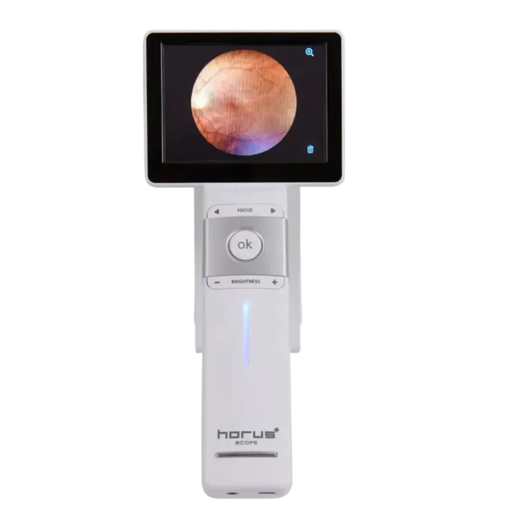
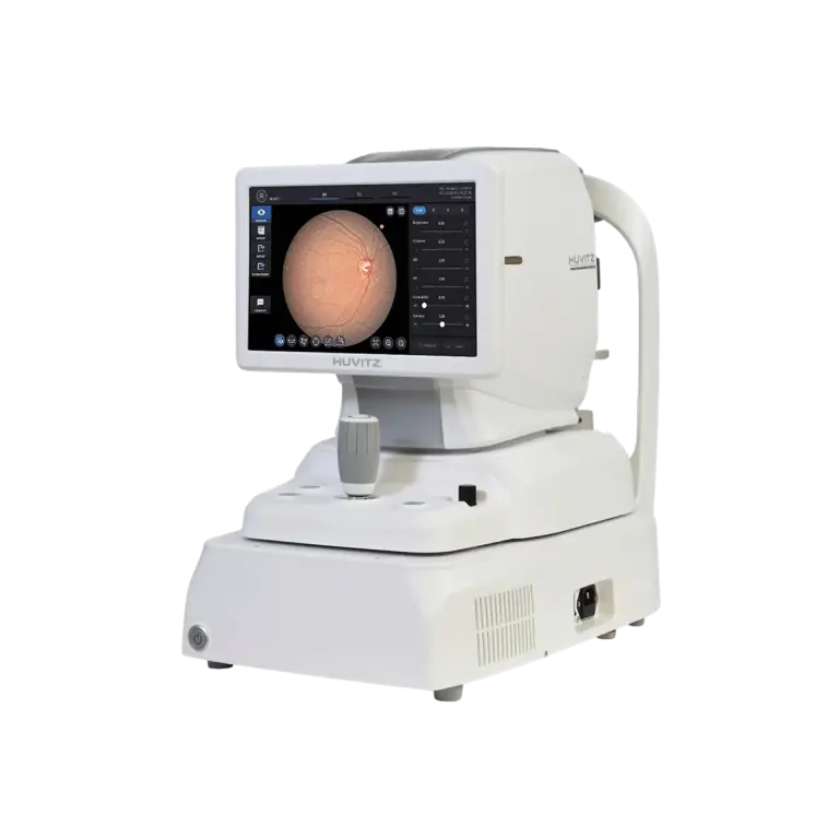
This page brings together the retinal cameras US Ophthalmic offers in the US: the fully automated Ezer EFC-2600 non-mydriatic camera, the Ezer Panorama ultra-widefield retinal camera, and the portable MiiS fundus camera for bedside and outreach imaging. US Ophthalmic is a US provider of ophthalmic equipment; we manufacture the Ezer line and back it with an in-house technical department and spare-parts stock. Tell us how you will use the camera and we will put together a quote.
How to choose a retinal camera
- Field of view: Standard cameras image the central retina in one shot; ultra-widefield systems capture much more of the periphery in a single image or montage. Match the field to whether you mainly screen the posterior pole or follow peripheral findings.
- Mydriatic or non-mydriatic: Non-mydriatic cameras image through an undilated pupil and suit screening workflows. Check the minimum pupil size each model needs.
- Automation: Automatic alignment, focus and capture let technicians take consistent images with little training and keep the patient's time at the camera short.
- Tabletop or portable: A tabletop camera gives the most consistent images in a fixed room. A portable fundus camera goes to patients who cannot sit at the instrument, such as in nursing homes, hospitals or outreach programs.
- Image management: Confirm how images are stored, exported and viewed, and that the software fits your EMR and network.
- Service and parts: Ask who services the instrument in the US, how consumables and spare parts are supplied, and what the US warranty covers.
Browse retinal imaging by type
Use the table to find the group that fits your practice, then open the category or model page for specifications.
| Type | Best for | Where to look |
| Automated non-mydriatic retinal cameras (Ezer EFC-2600) | Routine posterior-pole imaging and screening in a fixed room | This page |
| Ultra-widefield retinal cameras (Ezer Panorama) | Practices that follow peripheral retinal findings | This page |
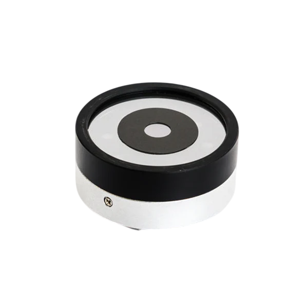
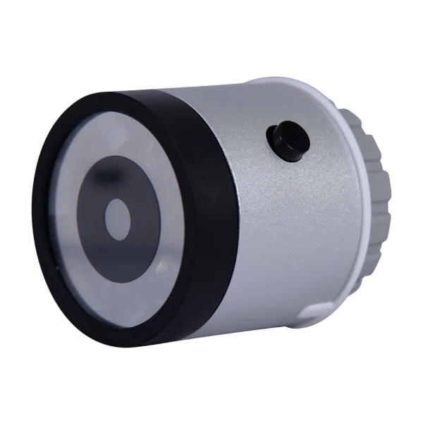
| Portable fundus cameras (MiiS) | Bedside, nursing-home and outreach imaging | Portable Retinal Cameras |
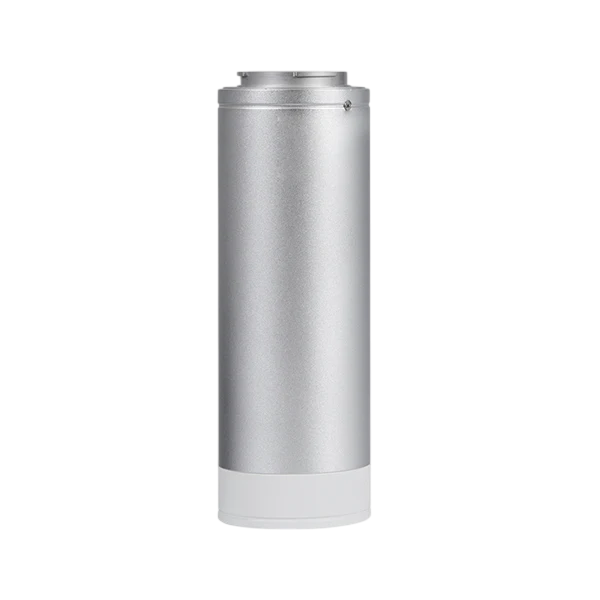
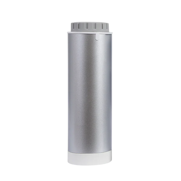
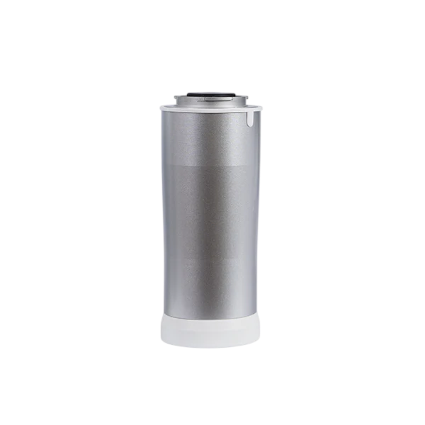
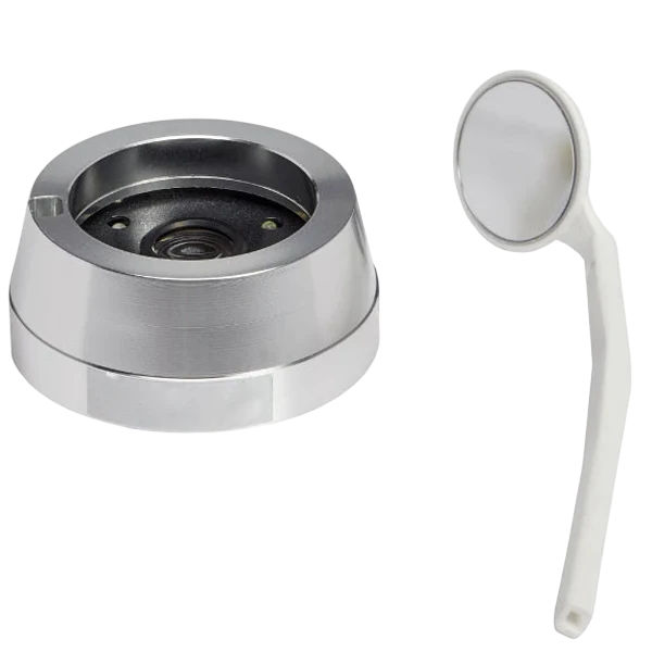
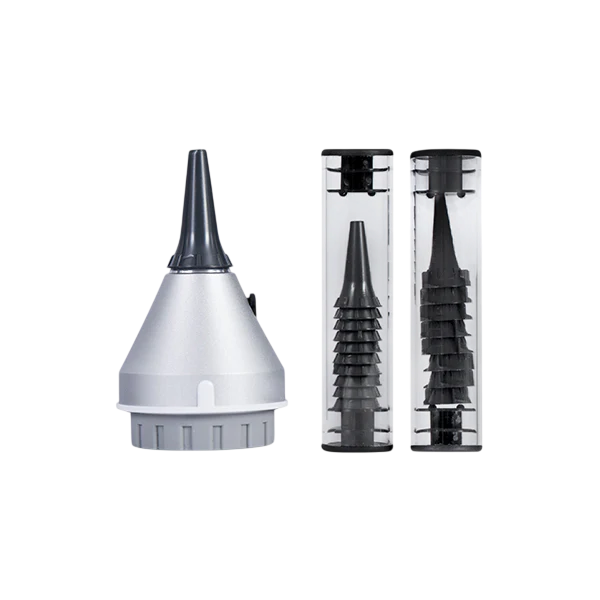
| Lenses and accessories | Extra lenses, cases and foot pedals for handheld imaging | Retinal Camera Lenses & Accessories |
Frequently asked questions
What does a retinal camera do? It photographs the retina, optic disc and macula so findings can be documented, compared over time and shared.
What is the difference between a standard and an ultra-widefield retinal camera? A standard camera images the central retina in one shot; an ultra-widefield camera captures a much larger area, including more of the periphery, in a single image or montage.
Do I need to dilate the patient? Not for a non-mydriatic camera, as long as the pupil meets the camera's minimum size; check the model page for that figure.
Do you sell refurbished retinal cameras? No. US Ophthalmic sells new equipment only. For a lower budget, contact us about open-box availability; open-box units are inspected and function-checked.
How do I get a quote? Request a quote with the models you are considering and how many lanes or rooms you are equipping, and we will come back with configurations, availability and lead times.
REQUEST QUOTE →