


-15%


Ezer Retinal Camera EFC-2600
The EFC-2600 is a top of the line fully automated non-mydriatic retinal camera with an advanced 3D tracking system capable of capturing high-definition retinal images in any eye care practice. The camera quickly delivers consistent colored ocular fundus images using multiple imaging modalities. The 45° field of view images help streamline screening of retinal abnormalities and early detection of diabetic retinopathy, glaucoma, macular degeneration and other growing disease. As easy-to-handle equipment, any eye care center can use the EFC-2600 to make a diagnosis without the need for a specialist. If an anomaly is detected, the telemedicine feature on the EFC-2600 is optimal for quickly obtaining the opinion of remote specialists.

Using the EFC-2600
The imaging process is not user dependent leading to high quality images regardless of the operator’s level of experience. The fully automated focus and capture features allow anyone to easily perform the examination and obtain the highest quality photos. The ease of imaging also allows for a relaxed and pleasant patient experience.

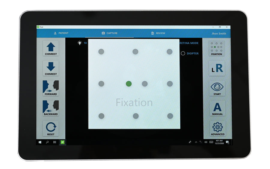
Live previews of the eye are visible on the touch screen. The operator begins the image capture with the press of just one button on the touch-screen and the software instructs the patient to focus on the green light while image capture occurs. The whole process is efficient and can take as little as 30 seconds per eye. The unintrusive nature of the EFC-2600 makes the whole imaging process quick and effortless for the patient and promotes high patient compliance.

Mosaic function — Enhanced image viewing and diagnosis
Retinal disease affects both the center and periphery of the retina. By providing an enhanced view of the retina mosaic fundus imaging has revolutionized the diagnosis of retinal disease. With ten selectable fixation targets, the mosaic modality of the EFC-260 allows the operator to take high-quality images of all points of interest on the retina up to 85 degrees.
The fully automatic mosaic function combines several retinal fields without user intervention to stitch together a seamless panoramic picture of the chosen fixation targets. These digital montages can be viewed on a large screen to facilitate the diagnosis of retinal pathologies.
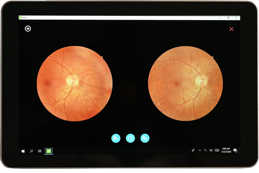
A Side by Side Comparison allows you to compare different images of the patient from different dates.

Easy image viewing and diagnosis
High resolution 12-megapixel camera and digital RED-Free images augment ability to view retinal vasculature allowing for quick diagnosis and detailed documentation of retinal changes associated with disease progression. Has a complement to RED-Free that allows you to view the RED-Free image in different gray scales.—The features, quality imaging and analysis provided by the EFC-2600 advance our current eye care to a future of high-quality, affordable healthcare for everyone.


RGB filters aid in the diagnostic process by increasing visibility of ocular disturbances. The red light is useful for imaging choroidal melanomas, choroidal nevi, pigmentary disturbances and choroidal ruptures and inflammatory lesions. The green light provides fine contrast and the greatest view of the fundus for observing normal and pathological retinal vascularization, hemorrhages and exudates. The blue light overcomes the barriers of white light to enhance the visibility of anterior retinal layers and provide detailed images of the retinal nerve fiber layer, inner limiting membrane, retinal folds, epiretinal membranes and cysts. The camera also has negative film that improves image contrast when light intensity is brighter.


Fundus imaging with the EFC-2600 enhances the accuracy and efficiency of early detection and appropriate diagnosis of retinopathies.
The ease of use and installation of the EFC-2600 allows any primary eye care center to operate and provide early detection of retinopathies reducing disparities across patient populations by improving access to screening.

The EFC-2600 is also equipped to take high quality images of the anterior segment, making it easy to capture images of the cornea, iris, ciliary body and lens allowing for early detection of anterior segment disorders.

Easy operation and pleasant patient experience
During examination the patient is instructed to rest their chin on the chin rest and forehead against the band. At this stage, the operator simply adjusts the motorized chin rest to the patient’s height through the intuitive interface using the large 10.1 inch color touch-screen display. The app interface allows the operator to select which eye to examine and select ten different internal fixation targets to image more fixation targets than other retinal cameras currently offer. The capture of the disc, fovea, macular or other peripheral images is thus easily enabled.


Rapid and early detection of diabetic retinopathy and other retinal diseases.
Diabetic retinopathy (DR) is the leading cause of permanent vision damage in adults. When left untreated, DR can lead to serious vision problems including vitreous hemorrhage, retinal detachment, glaucoma and ultimately blindness.

This incapacitating disease affects ~30% of adults over 40 with diabetes and is even more prevalent in underrepresented racial, ethnic and socioeconomic groups who do not have readily accessible screening. Although DR is so prevalent, patients may experience no symptoms at early stages, making early detection of DR critical. Low screening rates result in late referrals to specialists and vision loss.

Adaptability
The EFC-2600 has an adaptable interface and is easy to install in any eye care center. The smooth transfer of images is possible as the EFC-2600 is Digital Imaging Communications in Medicine (DICOM) compatible and easily integrates with other patient software.
Desktop Viewer
Patient's data can be transfered to de Desktop Viewer to be analized and storage separate Windows PC.
All electronic patient files and reports can be stored on the device and shared with specialists via cloud or email.


Increasing access to high quality healthcare through telemedicine
The EFC-2600 is much more than just a camera, it can generate dynamic reports. All electronic patient files and reports can be stored on the device and shared with specialists via cloud or email. It has output ports such as USB, which allows the exporting of patient images, and HDMI, which make it possible for images of the patient to be displayed on a larger screen.
These networking capabilities allow the device to support telemedicine services to provide expert opinions on all obtained images. The telemedicine feature provides reliable protection of patient data and improves access to exceptional quality healthcare. Specialists may view images and prepare medical reports 24 hours a day. This support cuts patient costs associated with visits to general practitioners and unnecessary hospitalizations.


AI
To aid the diagnostic process the EFC-2600 can implement artificial intelligence (A.I.) screening. The EFC-2600 can be integrated with any A.I. software. Recent advances in technology have trained A.I. to screen for vision threatening diseases with a high degree of accuracy.

As modern practices produce an overwhelming volume of images, assisted screening of images by A.I. is the future of efficient and objective ophthalmology. A.I. screening with the EFC-2600 streamlines diagnosis and maximizes efficiency at every practice.
Click here to see the brochure
Click here to see the manual
| Type | Digital non-mydriatic retina camera |
| Type of photography | Color, digital red-free, anterior eye image |
| Image Format | JPEG, PNG, Dicom (optional) |
| Field of View | 45 degrees |
| Minimal Pupil Size | 4.0 mm |
| Working Distance | 25 mm |
| Focus Adjustment Range |
-15D to +10D (w/o compensation lens) -30D to -10D or +5D to +30D (with compensation lens) |
| Flash Intensity | 10 levels, can be set manually |
| Light Source |
Observation light source : infrared LED Flash light source : White LED |
| Auto exposure | YES |
| Image | 12 MP |
| Eye Fixation | Internal ten points |
| Alignment | Fully automatic 3D tracking |
| Chinrest Movement | Motorized |
| Networking Capability | YES |
| Interface | USB 2.0, Ethernet, HDMI |
| Power Supply | AC 100V to 240V, 50/60Hz, auto selected |
| Operating Environment |
Temperature : 10℃ to 35℃ Humidity : 30% to 90% (no condensation) |
| Dimensions(W x D x H) | 282 mm x 485 mm x 492 mm |
| Weight | 38 pounds |
Primary Care
Click here to see the English Version
Eye Doctors
Click here to see the English Version
Atencion Primaria
Haz clic aquí para ver la versión en Español
Especialista
Haz clic aquí para ver la versión en Español
Back to Top

