












Skip to content
Search
+1 (888) 881-1122 REQUEST QUOTE

Clinical Areas:

Retina and Fundus

Glaucoma and Intraocular Pressure

Myopia Control

Cataract

Frey Perimeter AP-250

REQUEST QUOTEor call toll-free (888) 881-1122Financing available — simple terms, no leasing. Ask about it in your quote request.

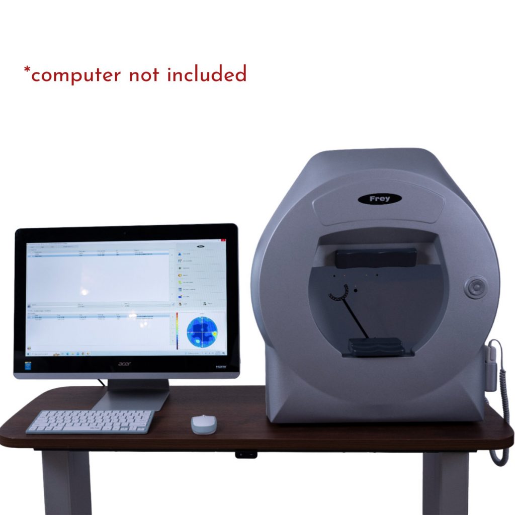
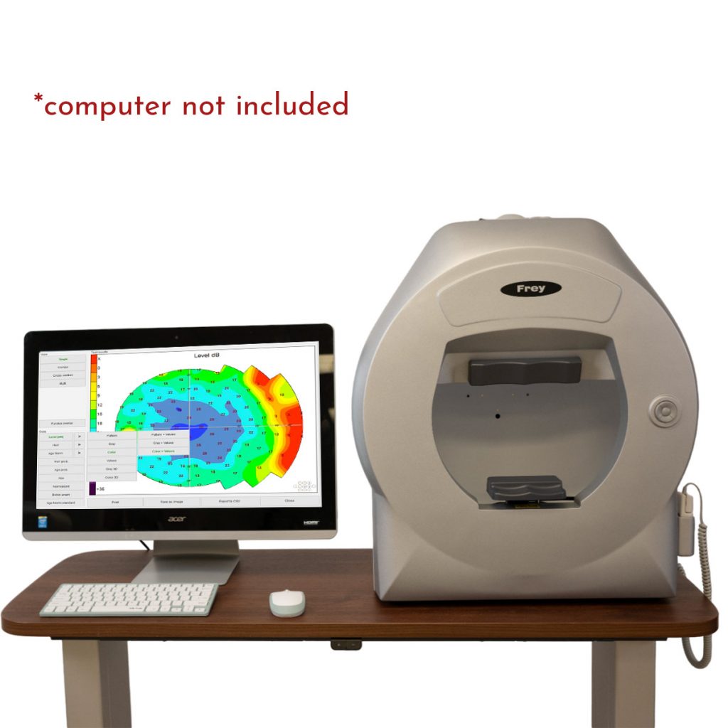
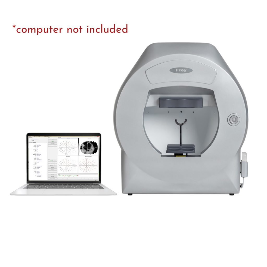
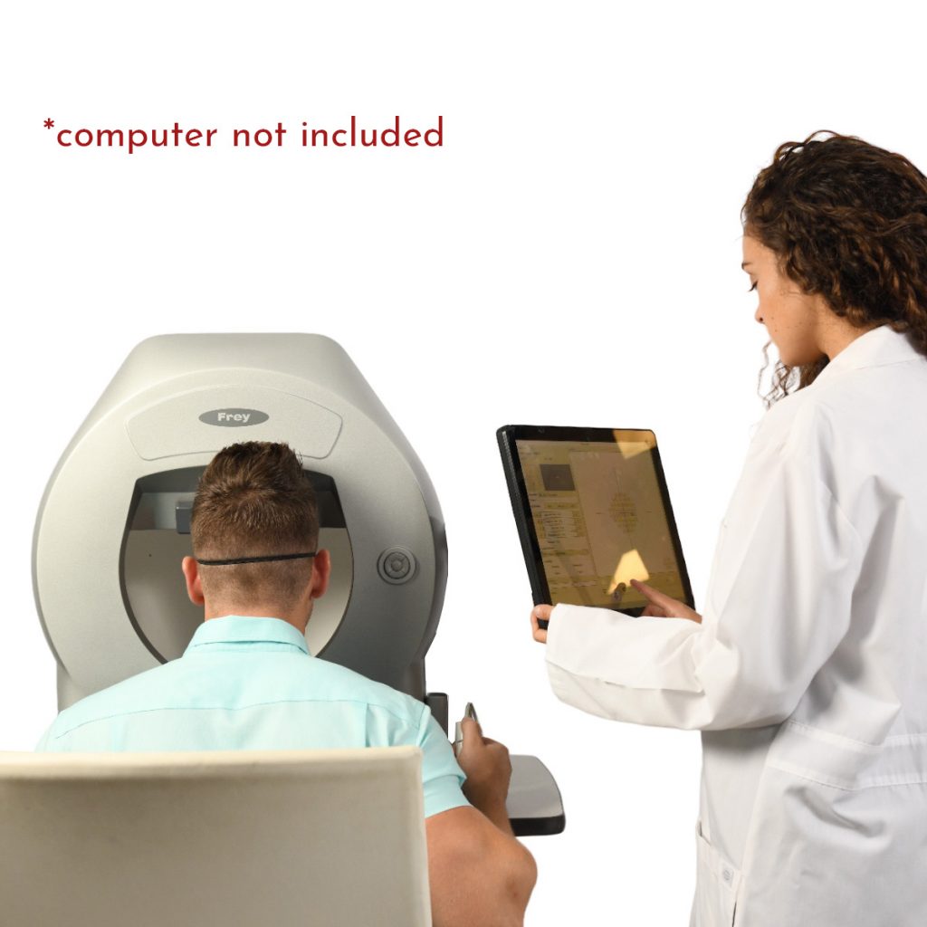
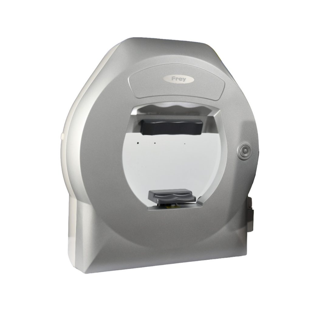
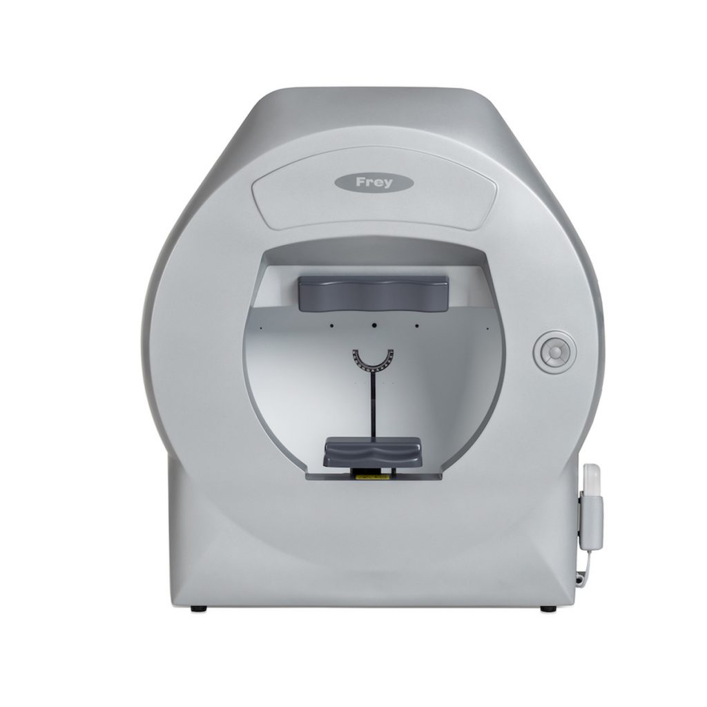
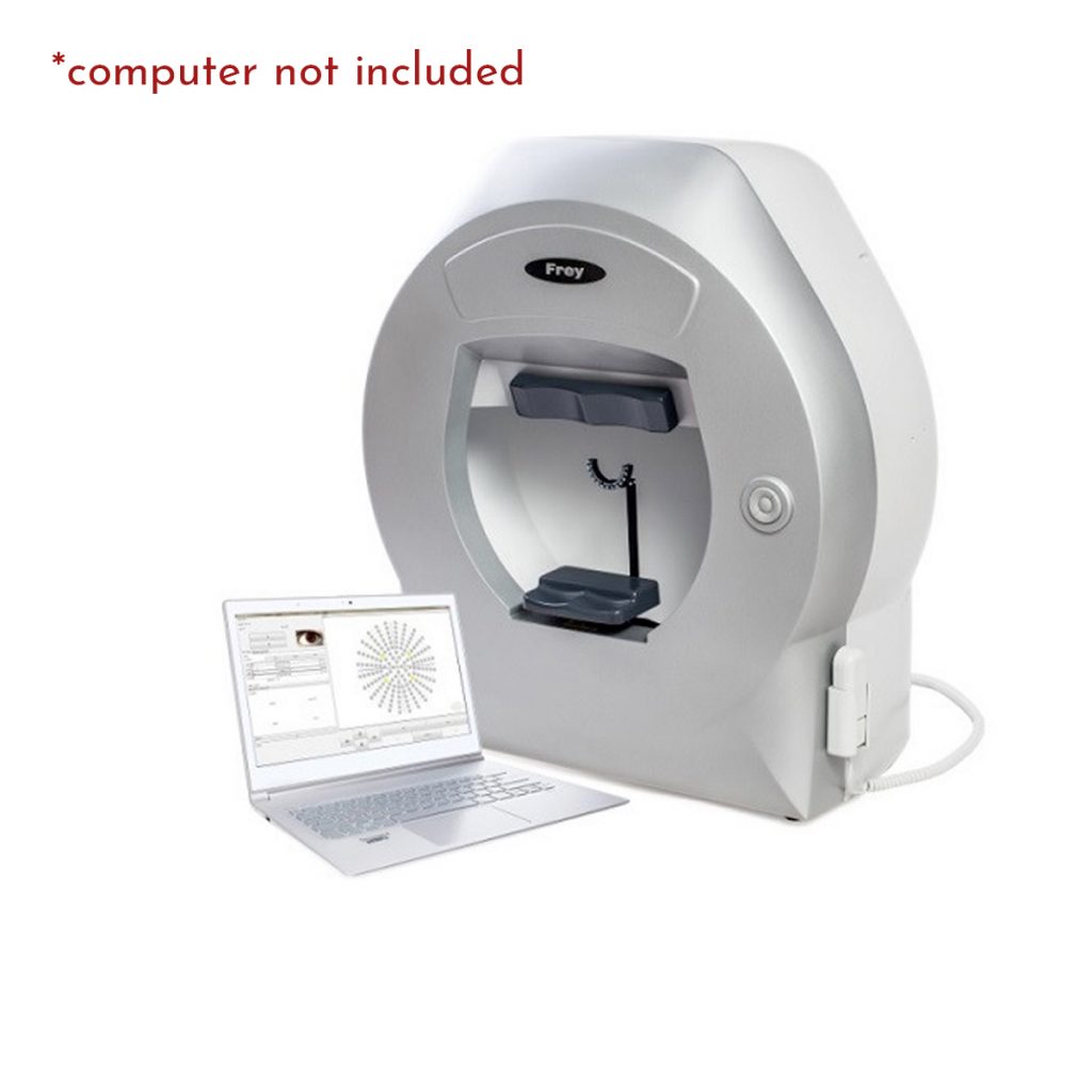
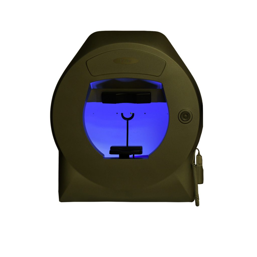
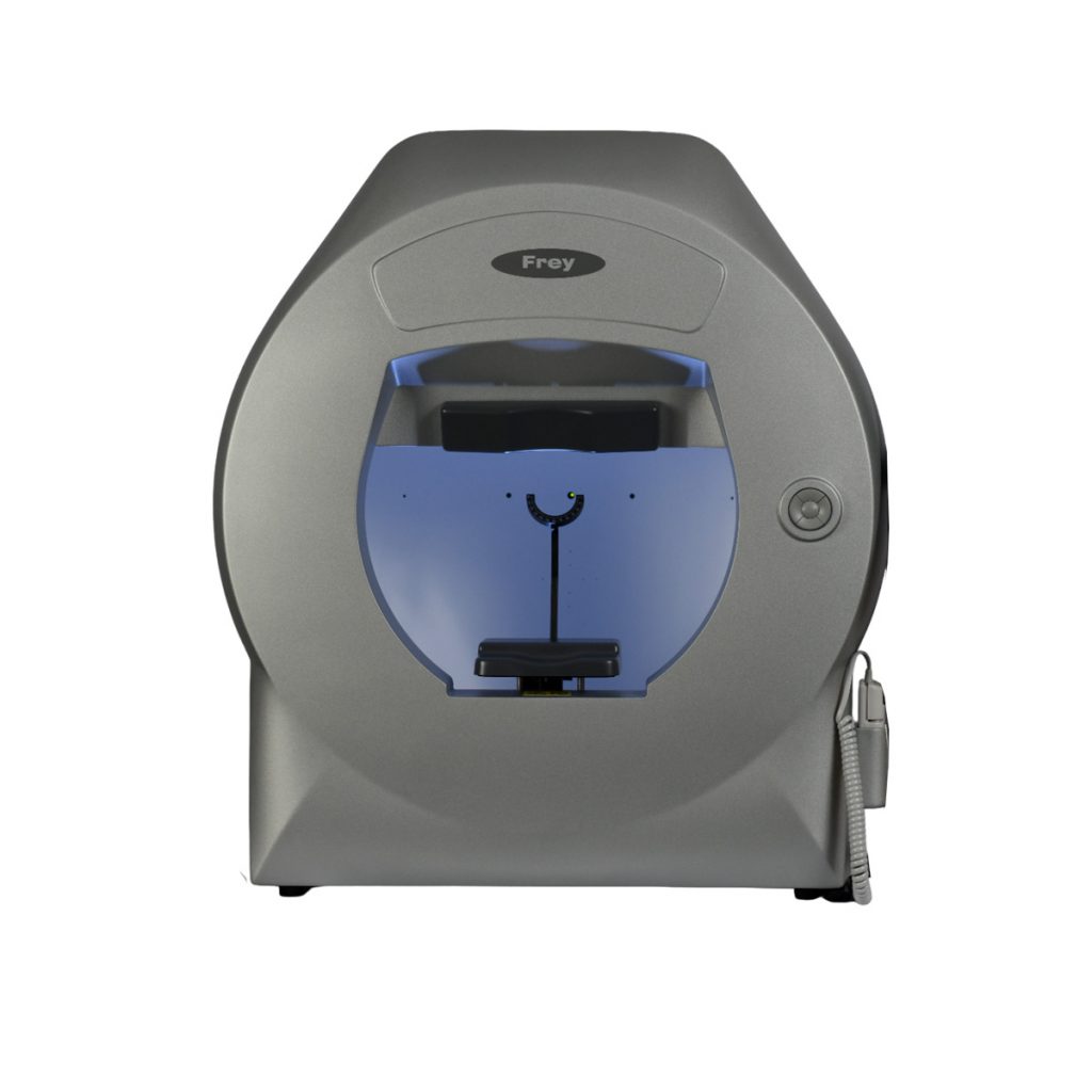
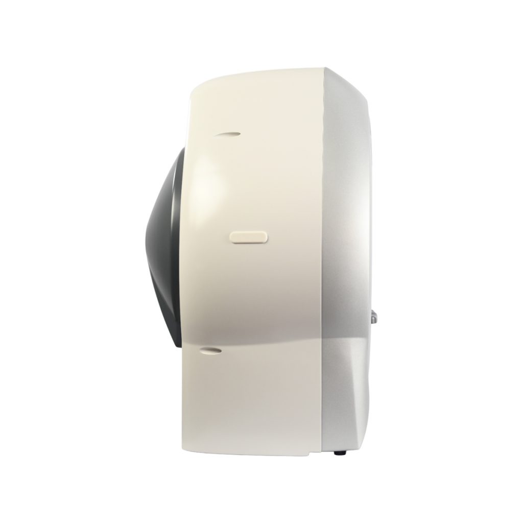
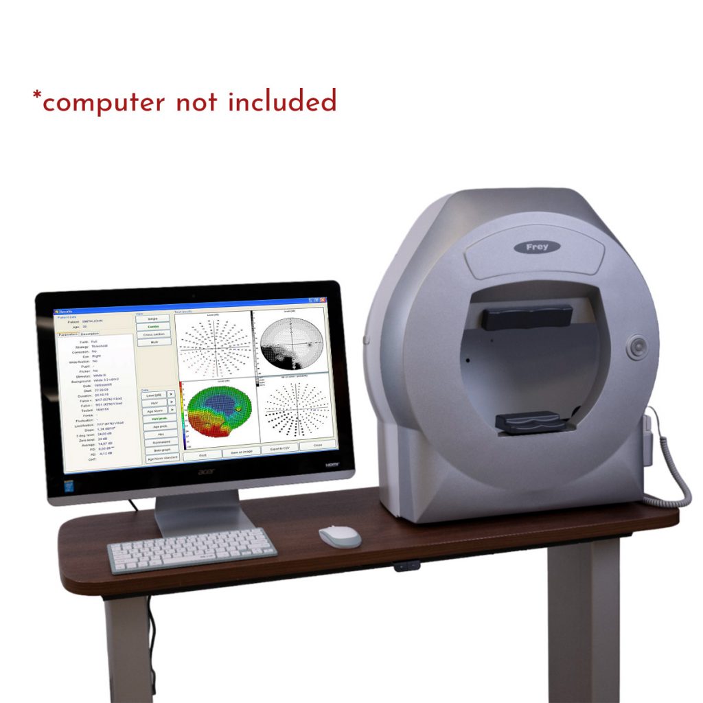
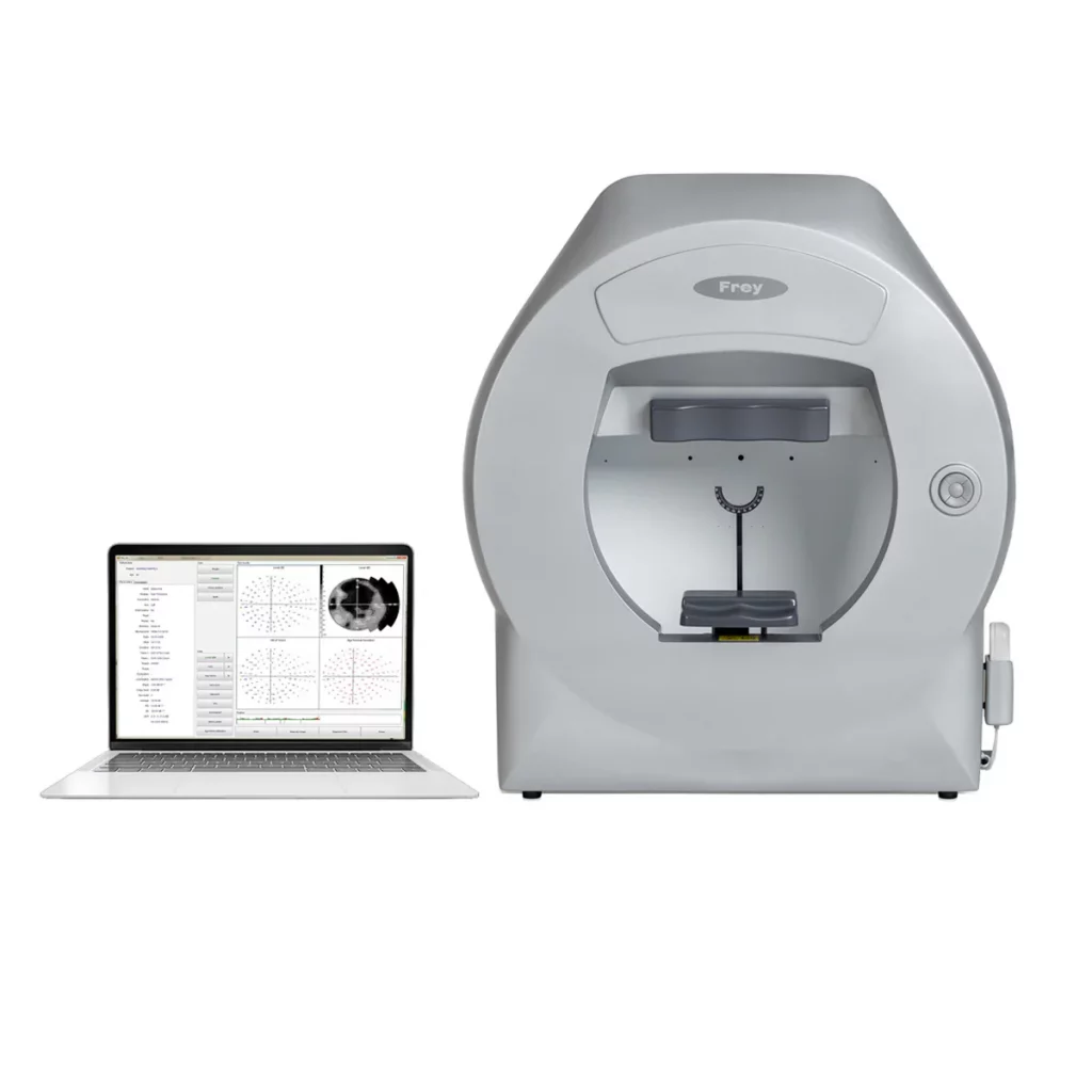
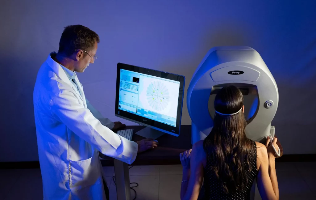
The AP-250 visual field analyzer is a fully functional static LED projection automated perimeter with full field measurement capability. The AP-250 uses green LED stimulus projection in Goldman size III. The software supplied with the device offers a wide variety of strategies, fields, and test parameters. Fixation control is performed automatically through the integrated camera or through blind spot position monitoring. Built-in data analyses include regression analysis and standardized formats for the presentation and printing of test results. The Frey AP-250 perimeter can be easily configured on any PC computer running the Windows operating system.

⚠ Monitor not included.
Frey Perimeter AP-250
Frey Perimeter AP-250 – image 24
Frey Perimeter AP-250 – image 25
Frey Perimeter AP-250 – image 26
Frey Perimeter AP-250 – image 27
Frey Perimeter AP-250 – image 28
Frey Perimeter AP-250 – image 29
Frey Perimeter AP-250 – image 30
Frey Perimeter AP-250 – image 31
Frey Perimeter AP-250 – image 32
Frey Perimeter AP-250 – image 33
Frey Perimeter AP-250 – image 34

Patient Focus – Greater Comfort

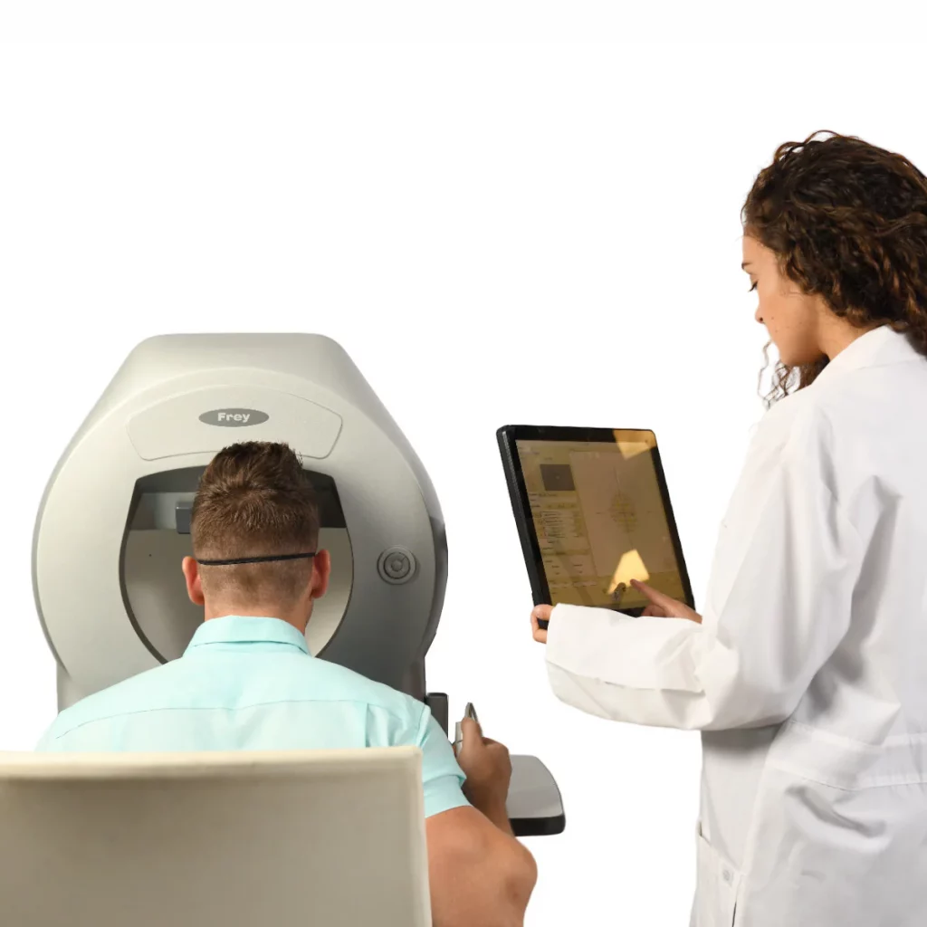
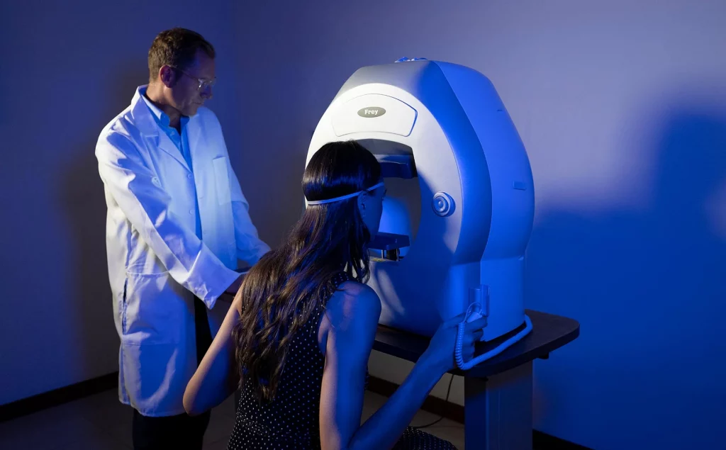
Visual field testing is a collaboration between the specialist and the patient. Also known as perimetry, it is a joint effort. A well-informed and comfortable patient generates greater reliability in the examination. This increased reliability translates into more precise and effective diagnosis and management by the physician. As a result, it improves clinical outcomes and patient satisfaction throughout the entire process, which can last decades. Frey offers greater comfort with a chin rest designed to provide stability. The patient’s head remains supported throughout the entire examination. The AP-250 features improved ventilation, reducing stress and increasing patient comfort.

Frey Perimeter AP-250 – image 1

Precise Results

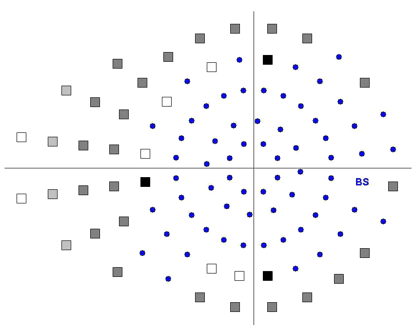
The stimulator dome offers a high density of concentric points. Advanced stimulus control, combined with automatic eye tracking, provides repeatable and precise examinations of the patient’s visual field loss.

Frey Perimeter AP-250 – image 2

Fast Examination Times

Efficient testing is performed using fast detection and threshold strategies, as well as enhanced fixation methods. Patients with advanced visual loss are assisted through pattern calibration and neurological methods.

Operating Modes:
Static Test Mode

The AP-250 emits fixed stimuli of variable luminance to determine the patient’s threshold. A dim light appears at a specific location and, if the patient does not perceive it, the intensity increases. The patient’s responses are compared to those of individuals from a control group of the same age.

The examination is defined by the Test and Strategy; all stimulus colors and sizes are available, and the results are more reliable and of higher quality.

Frey Perimeter AP-250 – image 3

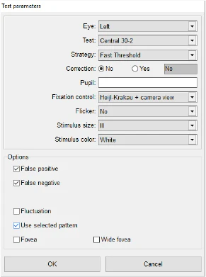
Static Test Parameters
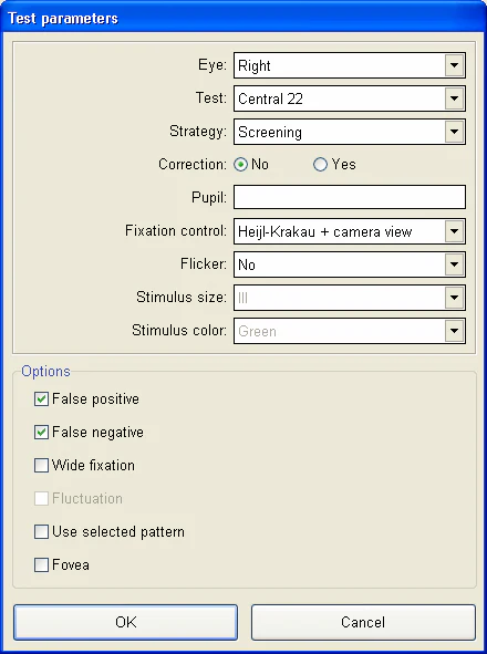
Two new parameters can be configured

  • Stimulus size
  • Stimulus color

The background color is white for all tests, except for the blue-on-yellow test. For that test, the stimulus size is Goldman III.

Selecting the appropriate test strategy is important, and with the AP-250> we test nearly twice as many points compared to competitors within the same visual field. Furthermore, the AP-250 provides fast scanning and intelligent thresholding, further enhancing examination accuracy.

Frey Perimeter AP-250 – image 4

Results Screen

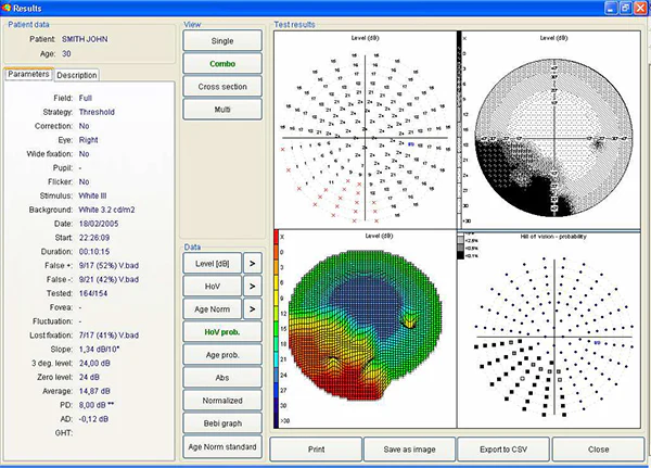
The “Results” screen is divided into functional panels. Detailed descriptions are provided in the following section (Individual, Combined, Cross-section and Multi).

Frey Perimeter AP-250 – image 5

Cross-Sectional View

It is a graphical map that divides the visual field into two parts represented by the colors red and blue. The study facilitates the selection of the cutting angle and allows sensitivity to be measured in decibels in all directions of the visual field up to 60˚.

Frey Perimeter AP-250 – image 6

Intuitive Software

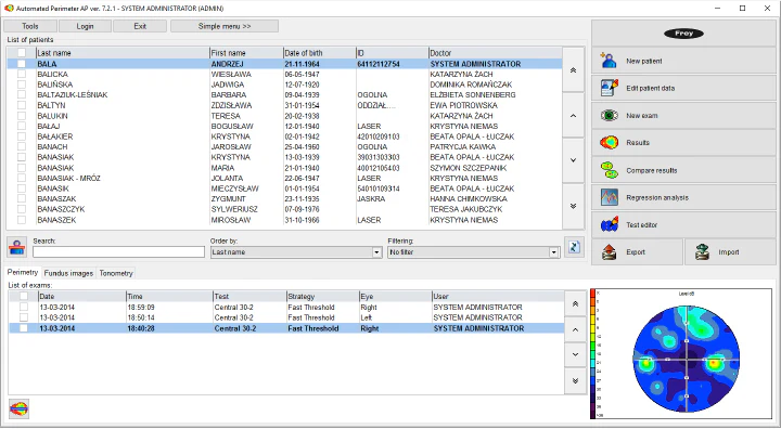
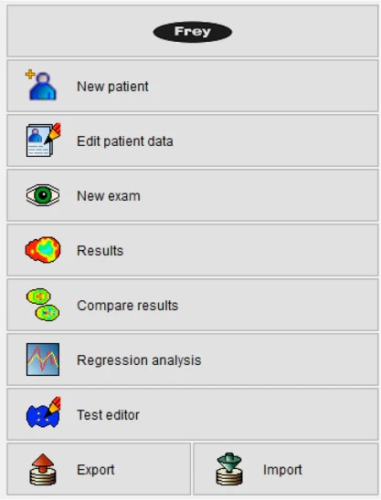
The design of the AP-250 is focused on both the patient and the clinical professional. The Frey perimeter software is intuitive and easy to use, enabling efficient and effective operation by a variety of qualified professionals. Interactive menus provide comprehensive information and efficient operation, reducing the time spent on exam preparation, review, and printing. This platform supports clinical excellence with increased patient throughput and greater efficacy.

  • Version information
  • Add new patient
  • Edit patient data
  • Start new exam
  • Results review
  • Comparison function
  • Regression analysis
  • Test editor
  • Export/Import function
Frey Perimeter AP-250 – image 7

Patient Image Bank

The perimeter application can be used as an image bank, for example, for fundus images.
DICOM fundus files can also be imported automatically.

Frey Perimeter AP-250 – image 8

Dual Home Screen Option

This feature allows two main screen options: a simplified one for use in the examination room by the operator, with personalized test selection and standardized procedures, and a full interface for reception areas or medical offices.

Frey Perimeter AP-250 – image 9

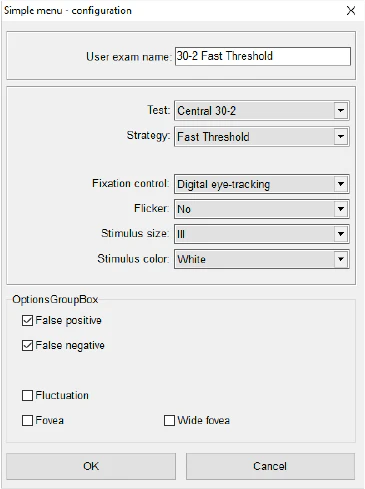
Simple Menu Customization

The simplified menu is fully customizable and all test parameters can be configured according to the customer’s request. The clinical workflow is also optimized, allowing standardization of the examination procedure. Furthermore, the software is easy to use and requires less prior knowledge.

Frey Perimeter AP-250 – image 10

Fixation Control

The AP-250 Automated Perimeter features two fixation control mechanisms.

Heijl-Krakau Method

A classic method that verifies the position of the blind spot by evaluating 11 random points to confirm correct eye positioning.

Gaze Position Method:
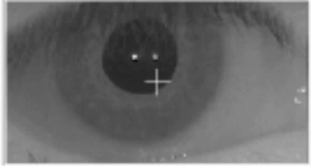
The AP-250 facilitates fixation analysis through a video camera that enables continuous pupil monitoring. The advantage is that it rejects patient responses when adequate fixation is not present.

Frey Perimeter AP-250 – image 11

Reliability

The automatic fixation control with integrated camera and eye tracking increases the repeatability and reliability of data capture. Automatic Pupil Detection If the automatic fixation monitoring method is used, the software detects the current position of the eye. It is not necessary to keep the eye centered on the image during the test, although it is essential that the patient always looks at the fixation point. The eye positioning control function can be accessed via the screen, the joystick, or the software. Additionally, the chin rest can be adjusted up/down and left/right.

Frey Perimeter AP-250 – image 12

Advanced Techniques

Previously, glaucoma was only diagnosed in advanced stages. Currently, new technologies enable the early detection of this condition.

Scintillation (Critical Flicker Fusion)

Glaucoma affects sensitivity to intermittent light stimuli.
CFF analyzes the patient’s ability to perceive changes between light and dark stimuli across different areas of the visual field.

It also measures early neuronal damage, detecting glaucoma in its initial stages.

Frey Perimeter AP-250 – image 13

Single Print

The AP-250 uses the simplest print format, which contains a large graphical map representing the examination results. It will print in the same format selected on the “Results” screen in “Single Mode”. When single mode is not selected, the dB level map in grayscale is printed by default. The advantage of the “single print” option is the high legibility of the printed report.

Frey Perimeter AP-250 – image 14

Multiple Printing Options

Users have the option to choose from 1 of 7 available print patterns. HFA-style printouts are predefined, as are standard static perimeter printouts (Frey, Medmont, etc.). Printouts can be produced in grayscale or in color.

Frey Perimeter AP-250 – image 15

Multiple Result Review Options

The AP-250 is equipped with a powerful set of data display modes.

Frey Perimeter AP-250 – image 16

Standard Calibration

The AP-250 was designed to reduce examination time in patients with significant visual field loss.
Previous examinations can be used as a starting point for a new test.
The “Standard” examination defines this starting point and, in addition, blind stimulus sites are evaluated at maximum intensity.
All test parameters are identical to the reference examination and are selected in the test parameters window when initiating the new examination.

Frey Perimeter AP-250 – image 17

Multiple Test Pattern Options

5 different test programs. Visual monitoring of both eyes of the patient.

Frey Perimeter AP-250 – image 18

LED LIGHT SOURCE

  • All light sources in the AP-250 are LED
  • The system requires no maintenance
  • Demonstrates high light source stability over time
  • True white-on-white perimeter
  • White stimulus with a color temperature of 4000K
  • White background illumination with a color temperature of 4000K
  • HFA devices use CCFL lamps for background illumination and a halogen bulb for the stimulus

Self-Diagnosis

Before and after each test, the AP-250 performs self-diagnosis cycles in which:

  • Stimulus intensity is verified by an internal sensor.
  • Any errors related to mechanical misalignment and light source intensity drift are minimized.
  • Dust or dirt accumulated in the optical path is detected.
  • Background illumination is stabilized to allow the test to begin.
  • The influence of ambient light is minimized.

Reliability

Reliable automated fixation control assists both the patient and the specialist through an integrated camera and automatic eye tracking. This increases the repeatability and reliability of data capture.

Regression Analysis – Static Perimetry

The AP software is equipped with options that allow observing changes in the patient’s vision in different ways. The first option is a simple comparison of results. The second, more advanced option is regression analysis, which helps observe the progression or regression of vision over time.
At least two examinations are required to perform regression analysis. The examinations must be conducted using the same strategy but on different days.
The patient’s name, age, and examined eye are displayed in the upper left corner of the screen. A selection box at the top center allows choosing one of five parameters to calculate and display the regression graph. Available parameters:

Decibel level
Decibel level of the Visual Hill
Age-corrected normal deviation (in decibels)
Pattern Deviation (PD)
Average Deviation (AD)

The selection box is available in “Individual Mode”. “Combined Mode” displays all five regression graphs together.

Frey Perimeter AP-250 – image 19

Probability Map of the Hill of Vision

A “probability map of the hill of vision” displays the probability of defects at each point of the visual field. Probabilities are calculated from the differences between the theoretical and measured hills of vision. The lower the probability, the greater the defect in the visual field.

Frey Perimeter AP-250 – image 20

Complete Analysis Modes

The reliability and functionality of the AP-250 are enhanced by complete analysis modes, supported by worldwide population statistics.
Shaded maps feature an improved 3D function.

Age-Normal Map

An age-specific normal range map displays the differences between age-normal values and the values obtained during the test. In this way, it reflects the difference between the theoretical range in dB and the measured value.
This map can be displayed in the following formats: numeric, grayscale, color scale, pattern scale, and 3D graph.

Frey Perimeter AP-250 – image 21

Multiple Testing Capabilities

Frey automated perimetry technology offers complete flexibility for both the specialist and the patient. This allows offering tests for glaucoma, full field, flicker, single binocular vision, and driving evaluation. This flexible platform provides visual field testing solutions covering a wide variety of clinical features.

Frey Perimeter AP-250 Frey Perimeter AP-250
Frey Perimeter AP-250 – image 22
Frey Perimeter AP-250 – image 23

Specifications

Frey Perimeter AP-250 specifications
Device Type Automated Perimeter
Measurement Dome Type Hemispherical, 300 mm radius with diffusing surface
Maximum Temporal Range (degrees) 80°
Stimulus Green on white, blue on yellow
Stimulus Duration 0.1 – 9.9s
Stimulus Size III
Stimulus Intensity 0.03 asb to 10,000 asb in 15 steps of 3 dB or 45 steps of 1 dB
Visual Field Test Distance 300 mm / 12 inches
Background illumination White 10 asb (3.2 cd/m²)
Fixation Control Heijl-Krakau blind spot monitor Eye tracking (video camera) Eye preview (video camera)
Test Models Age-corrected suprathreshold: Threshold detection: Full threshold, Fast threshold, Smart threshold. Others: 2 zones, 3 zones, defect quantification, constant, binocular, bi-conduction, directed perimetry, neurological.
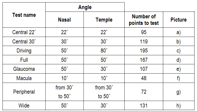
Library of Test Fields With concentric test point position settings: Macula, Central 22°, Central 30°, Full, Direction, Wide, Glaucoma, Peripheral. User-defined test fields: Bi-conduction, Occupational Medicine, Monocular, Binocular.
Correction Lens Diameter 38mm / 1.5 inches
Chin Rest Electric drive on both horizontal and vertical axes.
Computer Integrated PC touchscreen support
Printer External or network printer
Additional Software Functions Foveal threshold test, Automatic pupil measurement, User management module, Touchscreen operation, DICOM export, Network connectivity, Programming interface for EMR systems, HFA device data import, Automatic backup.
Dimensions Height: 637 mm / 25 inches Width: 566 mm / 22 inches Depth: 420 mm / 16.5 inches
Weight 18kg / 39.6lbs
Power Requirements Voltage 110–230 VAC Power 95 W

Manuals & Catalogs

Catalogs

Frey Perimeter AP-250

Frey Perimeter AP-250 – catalog cover
Catalogs

Frey Perimeter AP-250

Frey Perimeter AP-250 – catalog cover
Manuals

Frey Perimeter AP-250

Frey Perimeter AP-250 – manual cover