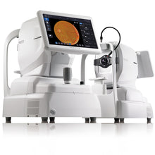
Huvitz Huvitz Med® - Fundus AI™
Huvitz Med® - Fundus AI™ provides major findings necessary for the diagnosis of retinal diseases based on fundus images.
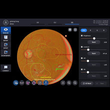
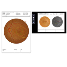
The AI solution provides major findings for the diagnosis of retinal diseases by locating abnormalities such as drusen, hemorrhage and hard exudate, detecting the abnormalities in fundus images and presenting patient reports.
In the current practice an ophthalmologist can directly examine the eyes by using special instruments such as schematic glasses, and dipole, and take a photograph of the fundus with a special camera for fundus examination.
In health screening centers, only preliminary screening is available, and patients are informed of the need for further examination through retina specialists (or ophthalmologists) for accurate diagnosis.
It takes extra time for examination with mydriasis. The accurate diagnosis may be difficult because it depends on retina specialists’ (or ophthalmologists) experience. In case of ophthalmologist with less experience, possible to miss some abnormal findings.


Huvitz Med® - Fundus AI™ helps intuitively check the presence of 12 abnormalities in different colors and locate them.
- Drusen
- Hemorrhage
- Hard Exudate
- Cotton Wool Patch
- Vascular Abnormality
- Glaucomatous Disc Change
- RNFL Defect
- Membrane
- Chroioretinal Atrophy
- Non-glaucomatous Disc Change
- Macular Hole
- Myelinated Nerve Fiber
Training Dataset
Based on a total of 103,262 fundus images on which 57 ophthalmologists* executed a triple reading, the diagnostic supporting system detects abnormalities in fundus images.
* References. Development and Validation of Deep Learning Models for Screening Multiple Abnormal Findings in Retinal Fundus Images. S0161-6420(19)30374-4.

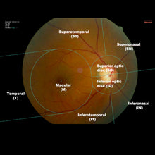
The software automatically locates optic discs and macula to mark eight regions of the fundus* and help diagnose the drawn regions.
- Macular
- Superior optic disc
- Inferior optic disc
- Temporal
- Superotemporal
- Inferotemporal
- Superonasal
- Inferonasal


