


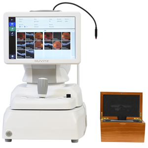


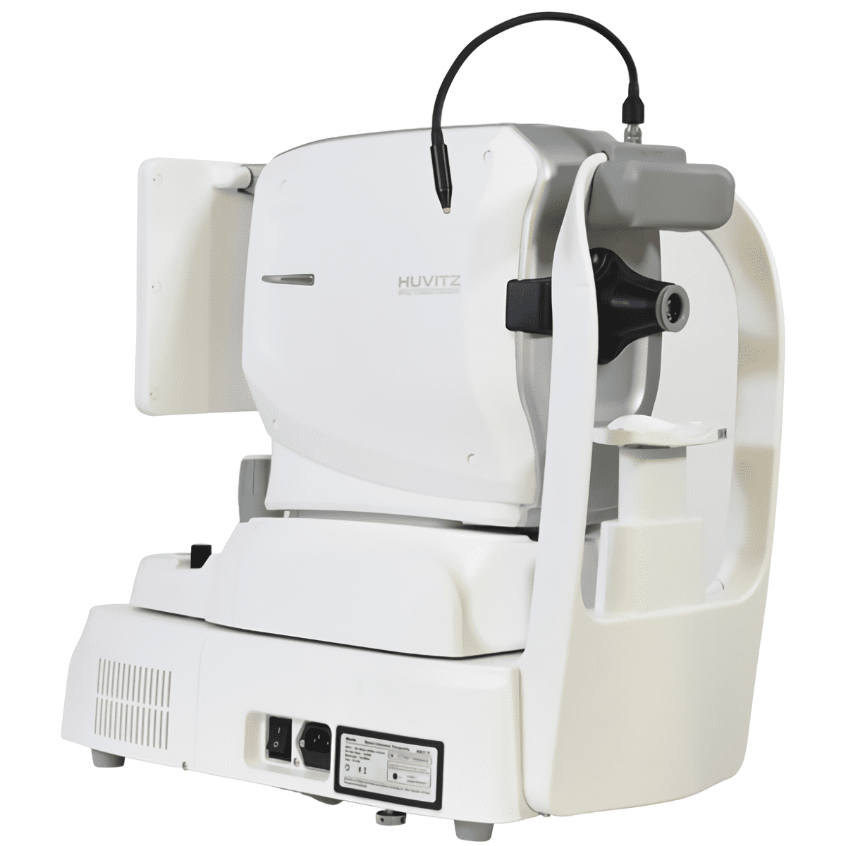
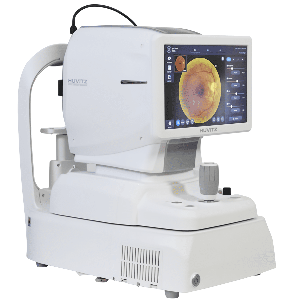
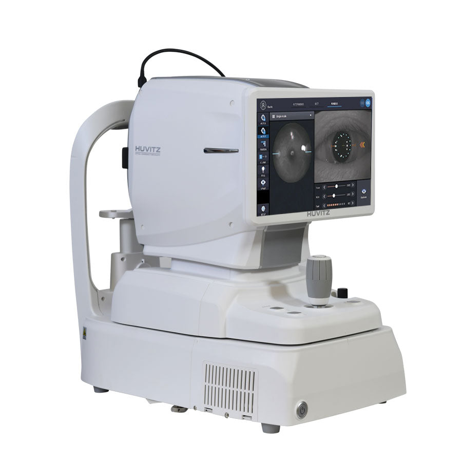
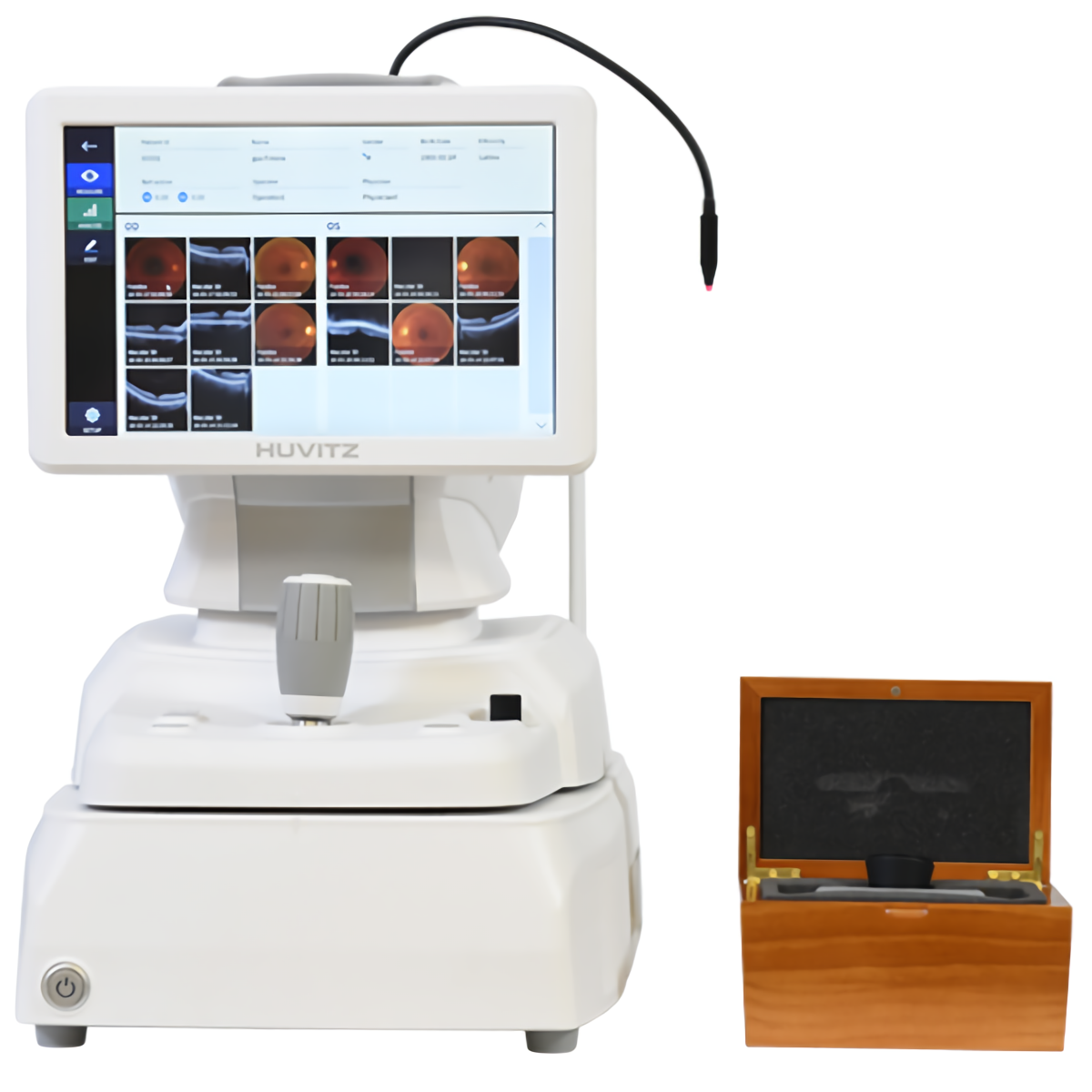
Huvitz Optical Coherence Tomography with Fundus HOCT-1F
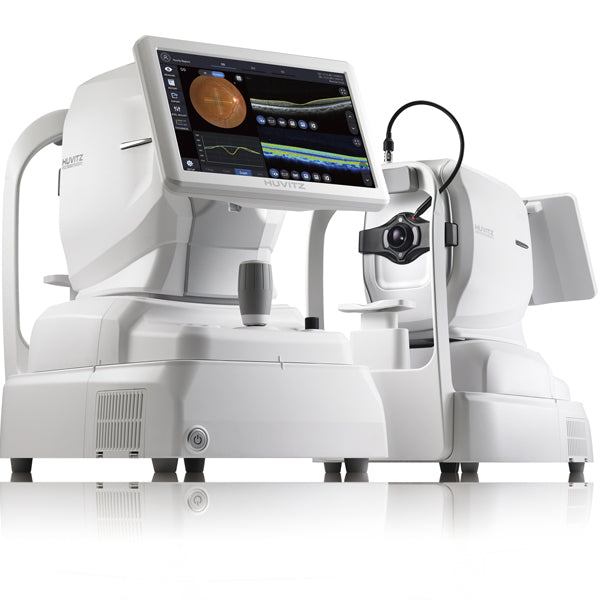
Everything in the HOCT is intelligent. 3D OCT & Fundus Camera is a fully integrated system combined with a PC. Provides OCT and Fundus data on a screen.
With how easy it is to use, with just one button, you can create a high-speed scanner and a high-quality image, which provides a greater perspective for the ophthalmology clinic. Easy to use and results are outstanding and easily follow. Huvitz All-in-One HOCT will be the icon for leading a new era of Optical Coherence Tomography(OCT).
 |
High Speed Provides High-speed Scan (68,000 A-scan/sec.), High-quality Image by using Huvitz’s outstanding optical technology and innovative image software. Shows extensive information, such as 3D structure of Retina, Macula's thickness and separation, in a vivid image. Accurate and Stable Image Averaging It is very important to obtain high-quality images that are accurate and stable in all OCTs. However, it’s not easy to capture these due to patient eye movement over the period of the test. The HOCT detects fast eye movements with image processing algorithms of fast Scan Speed* and Smart Viewing Technology(SVT)** and scans up to 68,000 points per second, and calibrates to create a high-quality optical image. HOCT can acquire high-quality images without any repetitive operation for first time users. *68,000 A-scan / sec., Less than 1.4 sec. in 6x6 mm2 3D shooting ** Smart Viewing Technology : Huvitz’s Speckle-Noise-Reduction System & Pre-Acquiring Algorithm to acquire high-quality images |
|||
 |
Vividly Visualized Retinal Layers Visualizing with precise B scans and smooth 3D images at faster scan speeds makes it easier to observe pathological shapes and status in stratified Retinal Layers. It is also useful to further elucidate the pathological rheobase of Macula and Optic Disc, including factors that impair Photoreceptor Function, Retinal & Choroidal Vasculature(vascular system) in a slice image for Retinal Layer consists of 7 pieces. |
|||
 |
Brightness Level Adjustment Precisely identify lesions by minutely adjusting image's brightness and contrast. In this way, specific parts of lesions can be highlighted which help users to easily see details. |
|||
 |
Combined-One By combining OCT, and PC, it can generate high resolution images providing multi-purpose functions for diagnosis. It saves both time and space by performing frontal view(Enface) of eye diseases, Tomography, cross-compare and diagnosis in a single run. Provides maximum psychological stability to the patient without re-shooting and reduces stress during shooting*. Easily checking lesion’s position by Fundus Image, it precisely guides the location of the OCT Scan Image. *Motion detection technology:Smart Scan Technology (SST) is applied to achieve perfect images without re-shooting even though there’re flicker or movement (see Smart Scan page). |
|||
|
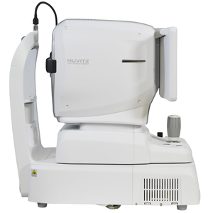
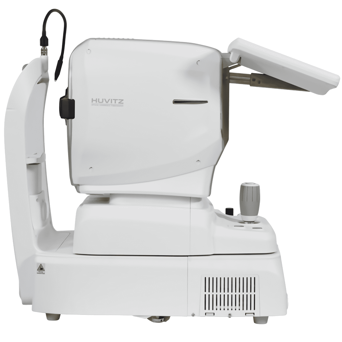
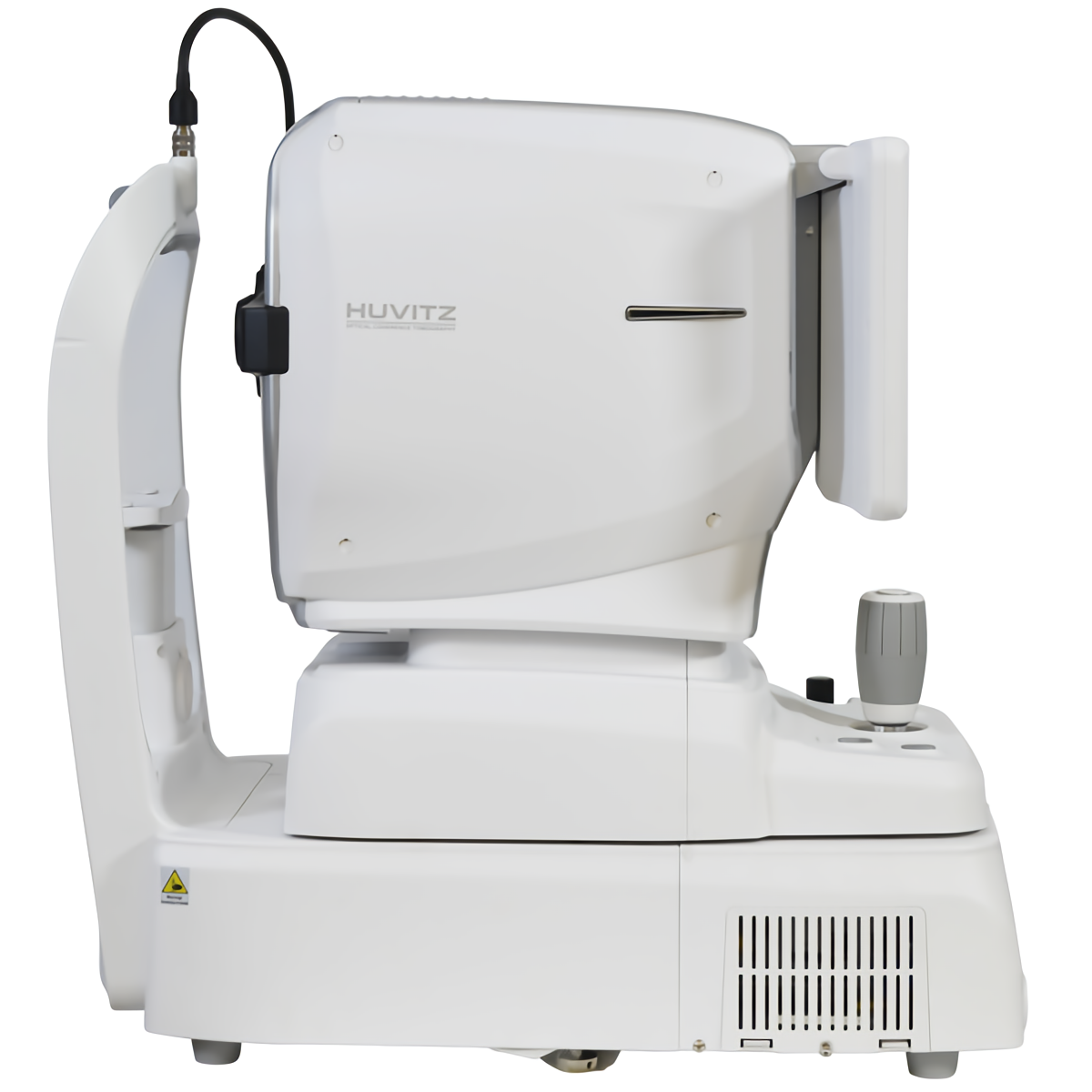
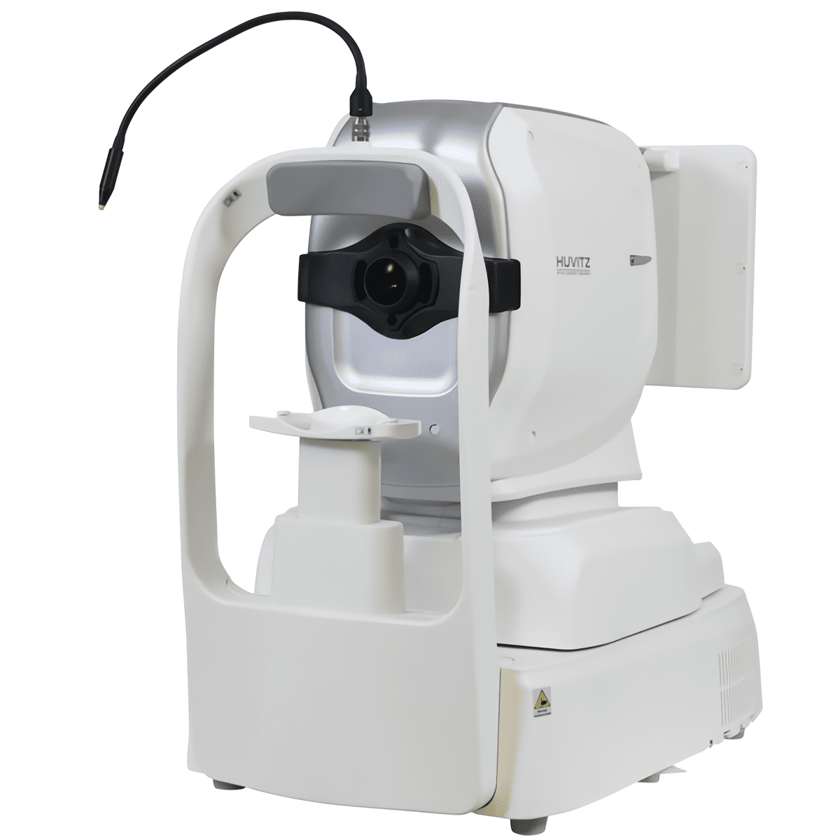
Compact Design Thanks to HOCT's space-saving design its perfect for hospitals and research areas with many diagnosis devices and treatment equipment, It can maximize the convenience of users as well as patients, thus saving time and space. |
||||
 |
Web Browsing System Patient's test data can be analyzed anywhere on the Internet. You can check and analyze all data of HOCT through Web Browser such as Internet Explorer, Safari, Chrome without installing special software separately. |
|||
|
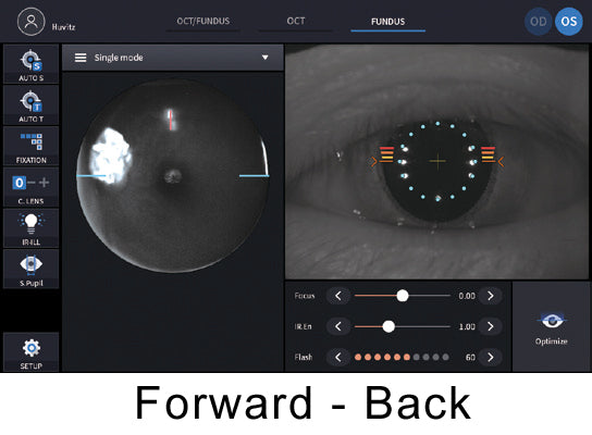
Fast and Stable Full Auto Mode Simply press the button once to capture the image quickly and easily without any errors with Auto Tracking, Optimize, Auto Shooting at the correct position. Depending on the application, select Semi Auto to obtain more detailed images. Semi Auto Mode You can obtain a more precise image by shooting Semi Auto Mode turning one’s gaze to the side for patients with eye diseases such as cataract, strabismus, or optic disk and peripheral measurements. Semi Auto Mode can also be applied to eyes with weak signals. XY alignment, focus is automatically adjusted, and manual operation during auto adjustment is also possible. Focusing and Firing functions can be judged and involved by users so that users can obtain images in an intuitive way. |
|||

|
Wide Area Scan(12mm x 9mm) A quick scan covers Macula and Optic Disk areas extensively. By scanning around Optic Disc or Macula for patient's pathological status, you can check the Thickness Maps between RNFL(Retinal Nerve Fiber Layer), GCL(Ganglion Cell Layer) and RPE Layers. Smart Scan Technology with Motion Detection Technology Image analyzer with Huvitz's unique Smart Scan Technology(SST) obtains a complete and perfect C-scan image by detecting any motion of eye flicker or movement that would prevent disappearance of scan line and image collection during measurement. |
|||
 |
Providing Various and Useful Scan Patterns 12 different patterns make it available to choose and apply the optimized pattern to the main symptoms or the area of retinal disease without repetitive work or time-wasting. |
|||
 |
Progression to track pathological changes OCT scan and fundus image of a patient can be compared at a glance to sequential measurement results from baseline to present. Progression from past to present helps analyze disease progression and treatment process. Thickness, Enface, and ETDRS can be superimposed on the IR or Fundus at each measurement point so that the change in thickness of nerve fiber can be confirmed according to the transition. It also provides a trace graph so you can study at a glande. |
|||
|
Compare before and after patient's symptom You can compare and analyze the baseline data of a patient with the current data. |
||||
 |
3D Modeling in High Speed and Wide Area High-speed, wide area(12mm x 9mm) 3D images help you quickly and comprehensively understand the condition of the Retina. Also, layer thickness maps can be used from ILM to RPE, respectively and Morphological changes on the measured surface of the layers can also be visually confirmed. |
|||
 |
OU to Cross-Analyze Function of Binocular Provides comparative analysis for Macular Thickness, RNFL Thickness, ONH(Optic Nerve Head) of binocular. |
|||
 |
Summary : Monocular-Scan and OCT / Fundus image Provides a summary analysis of Macula retina, RNFL, ONH at a glance. Helps identify whether follow-up examinations are needed or not. Easy to explain the results to the patient after diagnosis. |
|||
|
Detailed Report Provides patient's pathological structure and relevant & important data in easy-to-read format and also can print out the report on analysis screen. Analysis results can be viewed via Web Browser and printed out with different types of reports. |
||||
|
||||
 |
9mm Wide Chamber View Measurement of ACA(Anterior Chamber Angle) between cornea and Iris allows diagnosis and management of angle-closure glaucoma patients. 9mm High Resolution Cornea Thickness Measurement The 9mm high-resolution Cornea Scan provides an objective view of the structure of the eyeball and displays a cross-sectional image of the measured corneal thickness. |
|||
 |
Corneal Thickness Map Corneal's irregularity, Thinnest point, etc. can be identified with a corneal thickness map to visualize the patient's corneal thickness at a glande. |
|||
 |
High Resolution and Performance 12 Megapixel Camera High performance camera with Motion Artifact Suppression Technique provides high resolution images and also its low intensity of flash, fast & quiet operation maximize measurement quality. |
|||
|
Auto-Detection Of Pupil Size and Auto Flash Level Function It accurately measures the pupil size and automatically adjusts the intensity of light according to pupil size. Even patients with small pupil size can be easily measured without switching mode. Selecting Small Pupil Mode, to be adjusted more intensive light for the small pupil size. |
||||
 |
Panorama Function for Wide Range of Peripherals Multiple built-in capture color fundus images at different positions and automatically stitch them to optimized total overview. By providing high-resolution images with minimal distortion, you can immediately see key information for a comprehensive assessment of patient’ eye. |
|||
 |
Fixation Target for Flexible Configuration Fixation target can be set on the display for fine adjustment of a specific part of the eyeball. |
|||
|
CLINIC EXAMS with HOCT-1F High-quality, high-resolution OCT and color fundus images from HOCT are extremely useful for analysis and clinical diagnosis as the pathologic structure and status of each layer is accurately observed and recorded. |
||||
 |
Vitreomacular Traction(VMT) Syndrome : 80-years old, female, OD Vitreomacular traction syndrome is a potentially visually significant disorder of the vitreoretinal interface characterized by an incomplete posterior vitreous detachment with the persistently adherent vitreous exerting tractional pull on the macula and resulting in morphologic alterations and consequent decline of visual function. |
|||
 |
Glaucoma : 51-years old, male, OD Glaucoma is a disease that damages your eye's optic nerve. The same symptoms are found at Thickness map, Fundus, TSNIT chart, Clock chart. |
|||
 |
Macular Hole(MH) : 63-years old, female, OS A macular hole is a retinal break commonly involving the fovea. |
|||
 |
Macular Degeneration(MD) : 94-years old, male, OD Age-related macular degeneration is a disease that blurs the sharp, central vision you need for straight-ahead activities. |
|||
 |
Epiretinal Membrane(ERM) : 62-years old, male, OD Epiretinal membrane is a disease of the eye in response to changes in the vitreous humor or more rarely, diabetes. |
|||
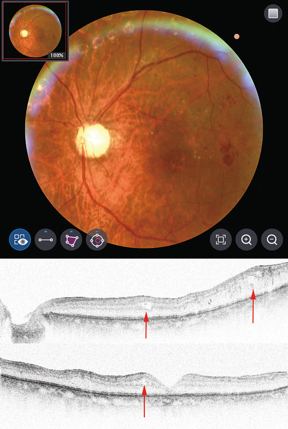 |
Diabetic Retinopath(DR) : 76-years old, male, OD Diabetic retinopathy is when high blood sugar levels cause damage to blood vessels in the retina. These blood vessels can swell and leak. Or they can close, stopping blood from passing through. Sometimes abnormal new blood vessels grow on the retina. |
|||
| Angio Module - Educational Video - Spanish Version |
Angiography (Optional Module) By One-Button, accurate details are provided with high-resolution images for vessels of retina & choroid and data of FAZ, flows, density. The function provides high-resolution images for vessels of retina & choroid with quantified index. It can be utilized with early diagnosis & treatment progression for macular degeneration, diabetic retinopathy, glaucoma, hypertensive retinopathy and retinal vein occlusion. Also, users can check abnormal vasculature in Custom View. Since the analysis shows index for FAZ, Flows, Density, it's easy to establish treatment plan. |
|||
 
|
* Detail Display for Accurate Index and Evaluation In Detail Mode, users can specifically observe vascular network per layer. Using analyzing tool, details of FAZ can be acquired conveniently. |
|||
 |
* Retina Layer Auto-Analysis The function provides high-resolution images for vessels of retina & choroid with quantified index. It can be utilized with early diagnosis & treatment progression for macular degeneration, diabetic retinopathy, glaucoma, hypertensive retinopathy and retinal vein occlusion. Also, users can check abnormal vasculature in Custom View. Since the analysis shows index for FAZ, Flows, Density, it's easy to establish treatment plan. |
|||
 |
* Detail Display for Accurate Index and Evaluation In Detail Mode, users can specifically observe vascular network per layer. Using analyzing tool, details of FAZ can be acquired conveniently. |
|||
 |
* Progression The Progression mode helps users to follow up the pathology of a disease. Binocular Comparison (OU) |
|||
 |
* In the Comparison Mode, users can check vascular network by layer in detail. By indicating layers in different colors, it's easy to check and understand the pathology of a disease. In case of comparison for diabetic retinopathy, the mode helps to track the pathology and establish treatment plan. |
|||
 * A Variety of Scan Sizes 3 mm / 4.5 mm / 6 mm / 9 mm HOCT-Angiography supports a variety of scan sizes, users can choose and observe per needs & cases. |
||||
 |
* Angio Panorama In case of checking Angiography image with large size, it is convenient to utilize the Angio Panorama function. *3 mm (Max. 12 mm x 9mm), 4.5 mm (Max. 13.5 mm x 9 mm) & manual mode are available |
|||
| Topography Module - Educational Video - Spanish Version |
Topography (Optional Module) Comprehensive Analysis by Topography in OCT Method : Providing Total Cornea Power Map able to measure Anterior & Posterior. Total Cornea Power Map Since the HOCT can analyze the posterior of the cornea, Users can now minimize errors caused by anterior & posterior axis of the cornea, corneal thickness, and refractive errors caused by the vitreous and corneal structures. |
|||
 
|
Compact Layout with Various Options With 16 Map types, Users can analyze Sim-K, Meridian, and Keratoconus data * Analysis Display / Numerical Information Users can analyze with either Single Eye, OU, or Comparison functionality. It is also possible to check for clinical changes after treatment or surgery with the Progression function. |
|||
  Because the Map displays accurate distance, size, and area with numerical information, the User is able to perform their analysis with confidence. Sim-K: Typical curve in order to utilize optimal IOL Lens calculation Meridian: Provide ø3, 5, 7 mm meridian by dividing the cornea into 3 areas Pachymetry: Total Thickness of Cornea Epithelium: Provide epithelium thickness at each point |
||||
 |
16 Map Types for Customized Treatment HOCT provides 16 map types including the anterior & posterior surfaces of the cornea. Users can display a wide variety of options to analyze and diagnose. In particular, posterior corneal surface measurements that allow for more accurate surgical outcomes.accuracy than the Placido or Scheimpflug methods. It minimizes motion artifacts because of its 2-second high speed capture rate. |
|||
  16 Map Types for Customized Treatment HOCT provides 16 map types including the anterior & posterior surfaces of the cornea. Users can display a wide variety of options to analyze and diagnose. In particular, posterior corneal surface measurements that allow for more accurate surgical outcomes. |
||||
| Biometry Module - Educational Video - Spanish Version |
Biometry (Optional Module) HOCT provides all the data you need for quick and easy calculations for optimizing the IOL lens power. Biometry from the Cornea to the Macula. From the Cornea to the Macula, HOCT displays 2D images and provides all data along the anterior and posterior segments. After measurement is complete, the User can identify and make adjustments where necessary. Also, it is possible to evaluate a dense cataract or defects in the macula. |
|||
 Visible Measurement by 2D Image User can easily adjust lines to analyze structures, by moving the cursor on the monitor in real-time. This can allow for a customized treatment of the non-typical Patient. |
||||
 More Precise Biometry Accurate Measurement by 2D Image: Visualize & Measure in Full Anterior Image of 16 mm * Confirmation Display able to Choose & Readjustment Users can make adjustments along the axial line, as well as remove measurements that fall outside of normal in order to create accurate statistics for the IOL calculation. |
||||
|
* Full Anterior Image for Wider Evaluation With the Wide Lens measurement, Users can obtain a full anterior image. With a simple click the CCT, ACD, ACA, W-to-W, LV, TISA, and TID* can be identified to aide in the diagnosis and management of Glaucoma. |
||||
|
* Contact Lens Fitting with Instant Check HOCT allows Users to check the suitability of Hard & Soft Contact Lenses for their Patients. It can also check the fit of an existing Contact Lens, quickly & precisely. |
||||
 |
* Reliable IOL Lens Recommendation HOCT recommends the optimal IOL Lens Power, based on biometry & corneal curvature with clinical data so that Users can easily make their surgical plan taking into consideration dense cataracts, corneal disease, or glaucoma. |
|||
  |
* View Function to Check the Basis of Disease Full Anterior Image, 12 types of Cornea Tomograph, Biometry Data (AL, LT, CCT, ACD) Tomography Image of Cornea. Comparison of asymmetry between left & right eyes with OU display. |
|||


Turn your OCT into an intelligent device with the installation of VUNO Med-Fundus AI

Click here to see the brochure
Click here to see the manual
| OCT | |
| Resolution(in Tissue) | Z:6~7um, XY:20um |
| A scan Rate | 68,000 A-scan/sec. |
| Scan Range | [Fundus] X:6-12mm, Y:6-9mm, Z:2.34mm [Cornea] X,Y:6-9mm |
| 3-D Acquisition Time | 1.4s(Fastest mode, A512 x B96) |
| Min. Pupil Diameter | Ø0.1in / 2.5mm |
| Light Source Wavelength | SLD 840nm |
| Optical Power at Cornea | ≤650uW |
| Scan Pattern | Macular Line, Macular Cross, Macular Radial, Macular Raster, Macular 3D, Disc Circle, Disc Radial, Disc Raster, Disc 3D |
| Fundus | |
| Camera | Color, Resolution 12MP |
| FOV | 45° |
| Min. Pupil Diameter | Normal:Ø 0.16in / 4mm / Small Pupil:Ø 0.13in / 3.3mm |
| Flash | LED |
| Resolution (on Fundus) |
Center:60 lines/mm or more Middle(r/2):40 lines/mm or more Middle(r):25 lines/mm or more |
| Common | |
| Working Distance | 1.3in / 33mm |
| LCD Size | 12.1"", Resolution 1280x800 |
| Dioptic Compensation |
Full Range:-33 to +33D -33 to -7D with Minus Compensation Lens+7 to +33D with No Compensation Lens |
| Fundus Surface Imaging | NIR/Enface, FOV: 40°x30° |
| Internal Fixation Lamp | LCD |
| Horizontal Movement | 70mm (back and forth) / 100mm (left and right) |
| Vertical Movement | 1.18in / 30mm |
| Chinrest Movement | 2.44in / 62mm, Motorized |
| Auto Alignment | X,Y for Positioning, Z for Working Distance |
| Auto Focusing | Diopter Adjustment for Focusing |
| Network | DICOM File support (Need to be customized) |
| Normative Database | Will be constructed after a release |
| Built in Computer | O |
| Others | |
| Power Supply | AC100 ~ 240V, 50/60Hz, 1.6A~0.7A |
| Dimensions / Mass | 13(W) x 21(D )x 20(H) in / 66lb (330(W) x 542(D )x 521(H) mm / 30kg) |
| Optional Accessories | Anterior Segment Adapter, Web Viewer |
|
Anterior Segment Module(optional) |
Scan Patterns: ACA Line, Cornea Radial, Cornea 3D Software Analysis: Corneal Layers, Thickness Map, Thickness & Angle |
| Web Viewer(optional) |
Web-based, Multy users can be accesible Progression Analysis, Comparion Analysis, 3D Analysis |
| OCT | |
| Resolution(in Tissue) | Z:6~7um, XY:20um |
| A scan Rate | 68,000 A-scan/sec. |
| Scan Range | [Fundus] X:6-12mm, Y:6-9mm, Z:2.34mm [Cornea] X,Y:6-9mm |
| 3-D Acquisition Time | 1.4s(Fastest mode, A512 x B96) |
| Min. Pupil Diameter | Ø0.1in / 2.5mm |
| Light Source Wavelength | SLD 840nm |
| Optical Power at Cornea | ≤650uW |
| Scan Pattern | Macular Line, Macular Cross, Macular Radial, Macular Raster, Macular 3D, Disc Circle, Disc Radial, Disc Raster, Disc 3D |
| Fundus | |
| Camera | Color, Resolution 12MP |
| FOV | 45° |
| Min. Pupil Diameter | Normal:Ø 0.16in / 4mm / Small Pupil:Ø 0.13in / 3.3mm |
| Flash | LED |
| Resolution (on Fundus) |
Center:60 lines/mm or more Middle(r/2):40 lines/mm or more Middle(r):25 lines/mm or more |
| Common | |
| Working Distance | 1.3in / 33mm |
| LCD Size | 12.1", Resolution 1280x800 |
| Dioptic Compensation |
Full Range:-33 to +33D -33 to -7D with Minus Compensation Lens+7 to +33D with No Compensation Lens |
| Fundus Surface Imaging | NIR/Enface, FOV: 40°x30° |
| Internal Fixation Lamp | LCD |
| Horizontal Movement | 70mm (back and forth) / 100mm (left and right) |
| Vertical Movement | 1.18in / 30mm |
| Chinrest Movement | 2.44in / 62mm, Motorized |
| Auto Alignment | X,Y for Positioning, Z for Working Distance |
| Auto Focusing | Diopter Adjustment for Focusing |
| Network | DICOM File support (Need to be customized) |
| Normative Database | Will be constructed after a release |
| Built in Computer | O |
| Others | |
| Power Supply | AC100 ~ 240V, 50/60Hz, 1.6A~0.7A |
| Dimensions / Mass | 13(W) x 21(D )x 20(H) in / 66lb (330(W) x 542(D )x 521(H) mm / 30kg) |
| Optional Accessories | Anterior Segment Adapter, Web Viewer |
|
Anterior Segment Module(optional) |
Scan Patterns: ACA Line, Cornea Radial, Cornea 3D Software Analysis: Corneal Layers, Thickness Map, Thickness & Angle |
| Web Viewer(optional) |
Web-based, Multy users can be accesible Progression Analysis, Comparion Analysis, 3D Analysis |
HOCT-1 - Step by Step - English Version
HOCT-1 - Step by Step - Spanish Version
HOCT-1 - Step by Step - Portuguese Version
HOCT-1F - Introduction to the Modules - English Version
HOCT-1F - Topography Module - English Version
HOCT-1F - Angiography Module - English Version
HOCT-1F - Biometry Module - English Version
HOCT-1F - Introduction to the Modules - Spanish Version
HOCT-1F - Topography Module - Spanish Version
HOCT-1F - Angiography Module - Spanish Version
HOCT-1F - Biometry Module - Spanish Version
HOCT-1F - Introduction to the Modules - Portuguese Version
HOCT-1F - Topography Module - Portuguese Version
HOCT-1F - Angiography Module - Portuguese Version
HOCT-1F - Biometry Module - Portuguese Version
HOCT-1F - Commercial Presentation - Spanish Version
HOCT-1F - Commercial Presentation - Portuguese Version
HOCT-1F - Commercial Presentation - Spanish Version
HOCT-1F - Commercial Presentation - Portuguese Version
HOCT-1F - Commercial Presentation - Spanish Version
HOCT-1F - Commercial Presentation - Portuguese Version


