AUTOMATIC DETECTION OF MEIBOMIAN GLANDS IN UPPER AND LOWER EYELIDS
Meibomian glands (MGs) play a significant role in tear quality by producing lipids (meibum) that form part of the superficial tear film. MG dysfunction destabilizes the tear composition, resulting in evaporative dry eye. The posterior lamella of the eyelid houses a set of parallel MGs located between the tarsal conjunctiva and the tarsal plate. A normal Meibomian gland is approximately linear and measures between 3 and 4 mm in length, traversing the posterior eyelid perpendicularly to the lid margin. A more detailed inspection of a Meibomian gland reveals a tubuloacinar architecture with saccular arrangements of acini and a ductal system that communicates with the orifices near the mucocutaneous junction of the eyelid. The glandular acini contain clusters of modified sebaceous cells called meibocytes (the functional unit of the Meibomian gland). These cells synthesize and secrete lipids (meibum) into the precorneal tear film. Meibum impregnates the tear surface, where it fulfills several important functions. It prevents tear evaporation and, consequently, desiccation of the ocular surface; it acts as a physical and hydrophobic barrier against the inward movement of environmental and organic agents; and it lubricates the ocular surface to prevent irritation, while simultaneously promoting a clear ocular image. Accordingly, tear physiology depends on the proper functioning of the MGs.
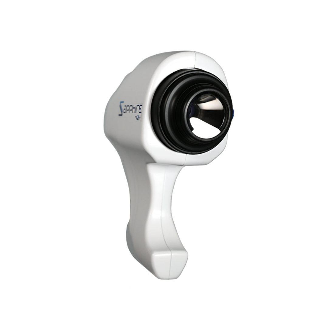
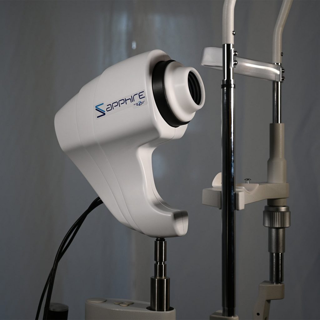
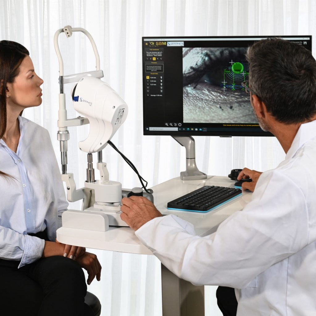
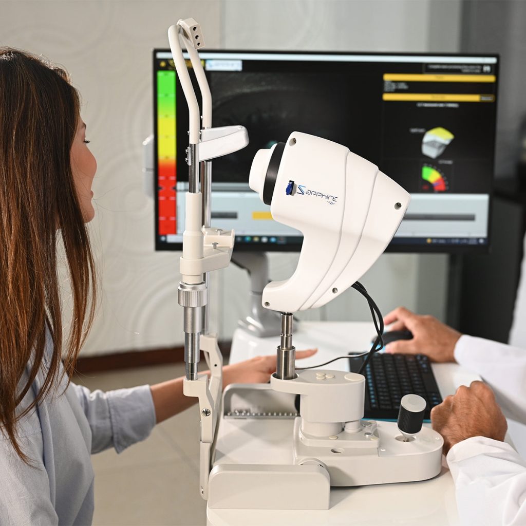
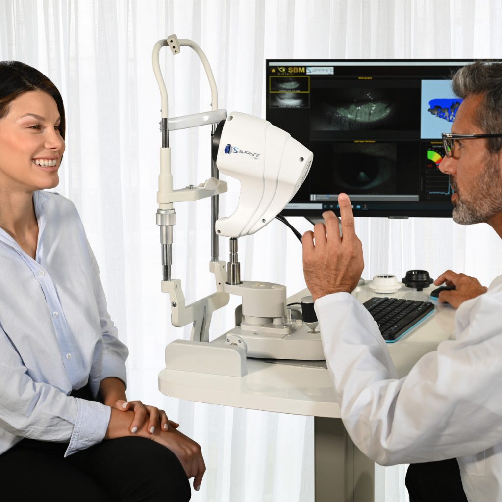
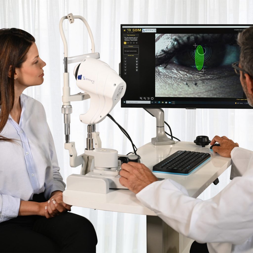
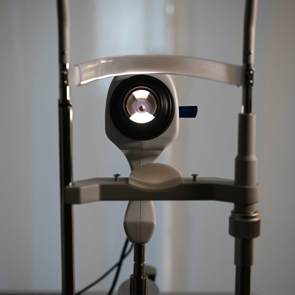
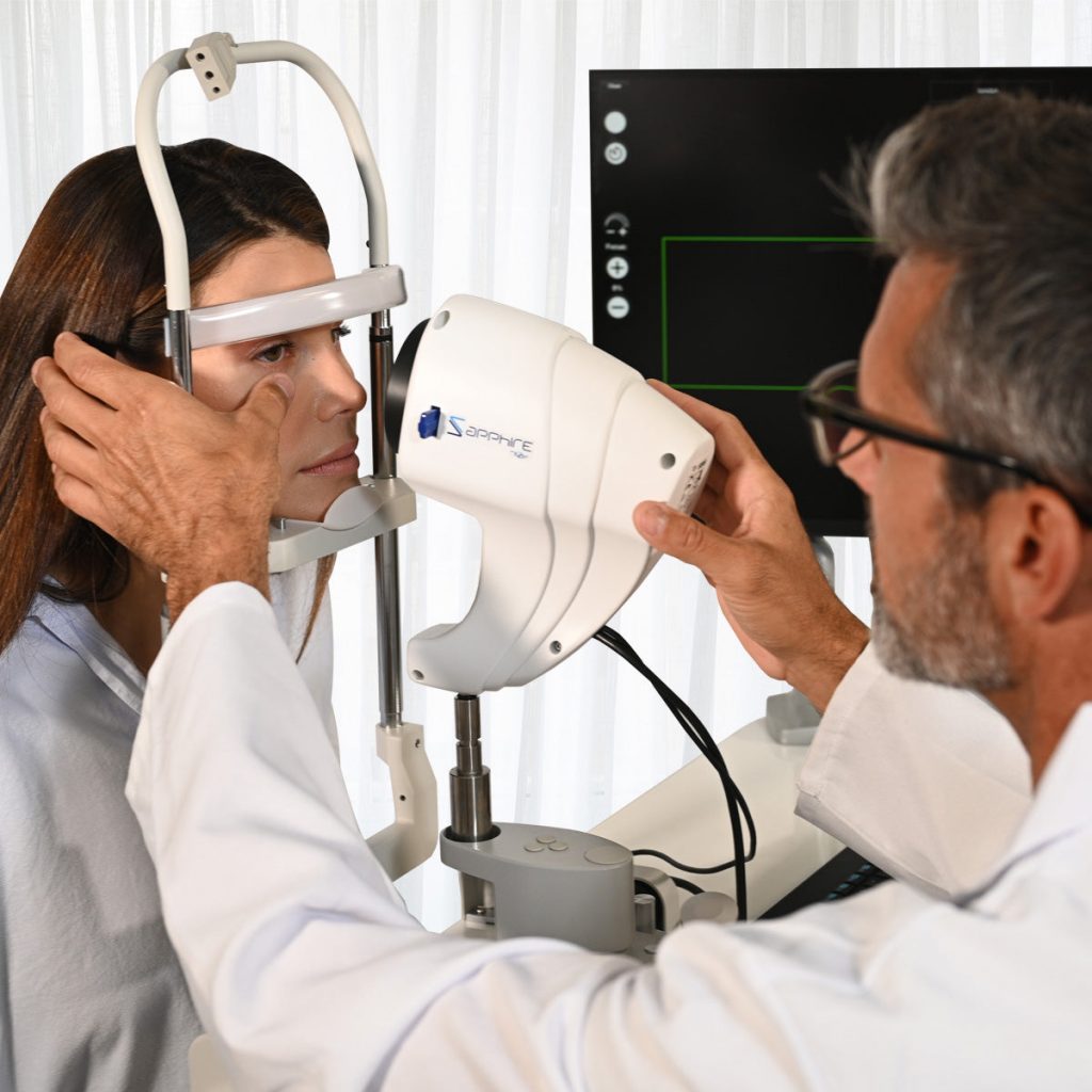
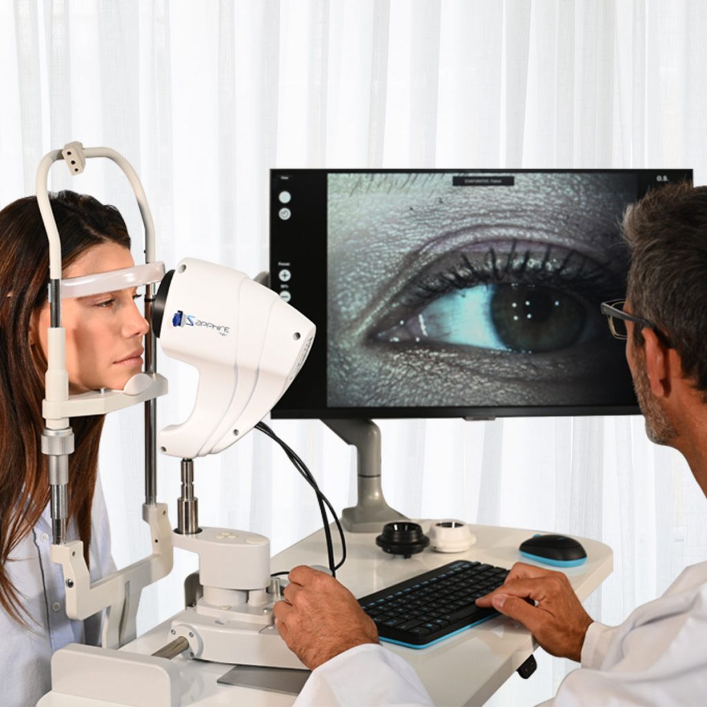
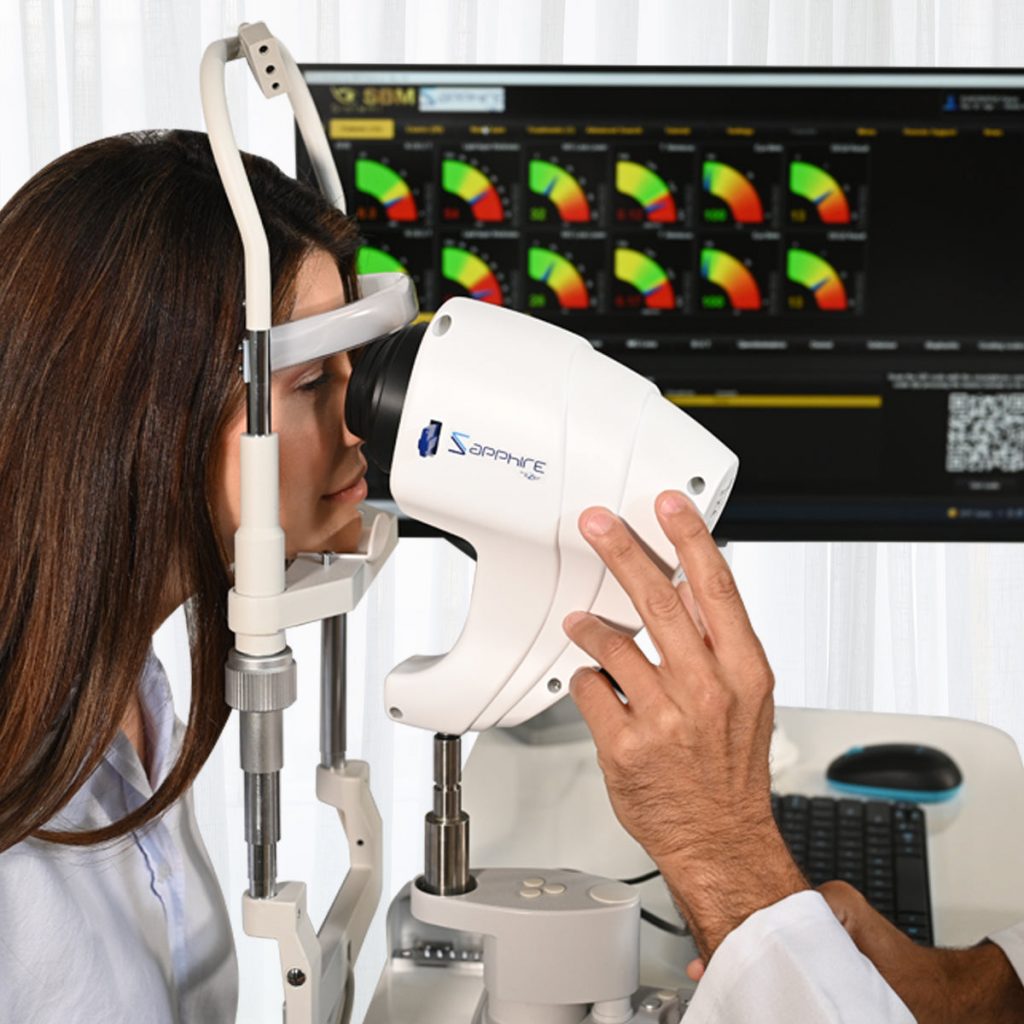
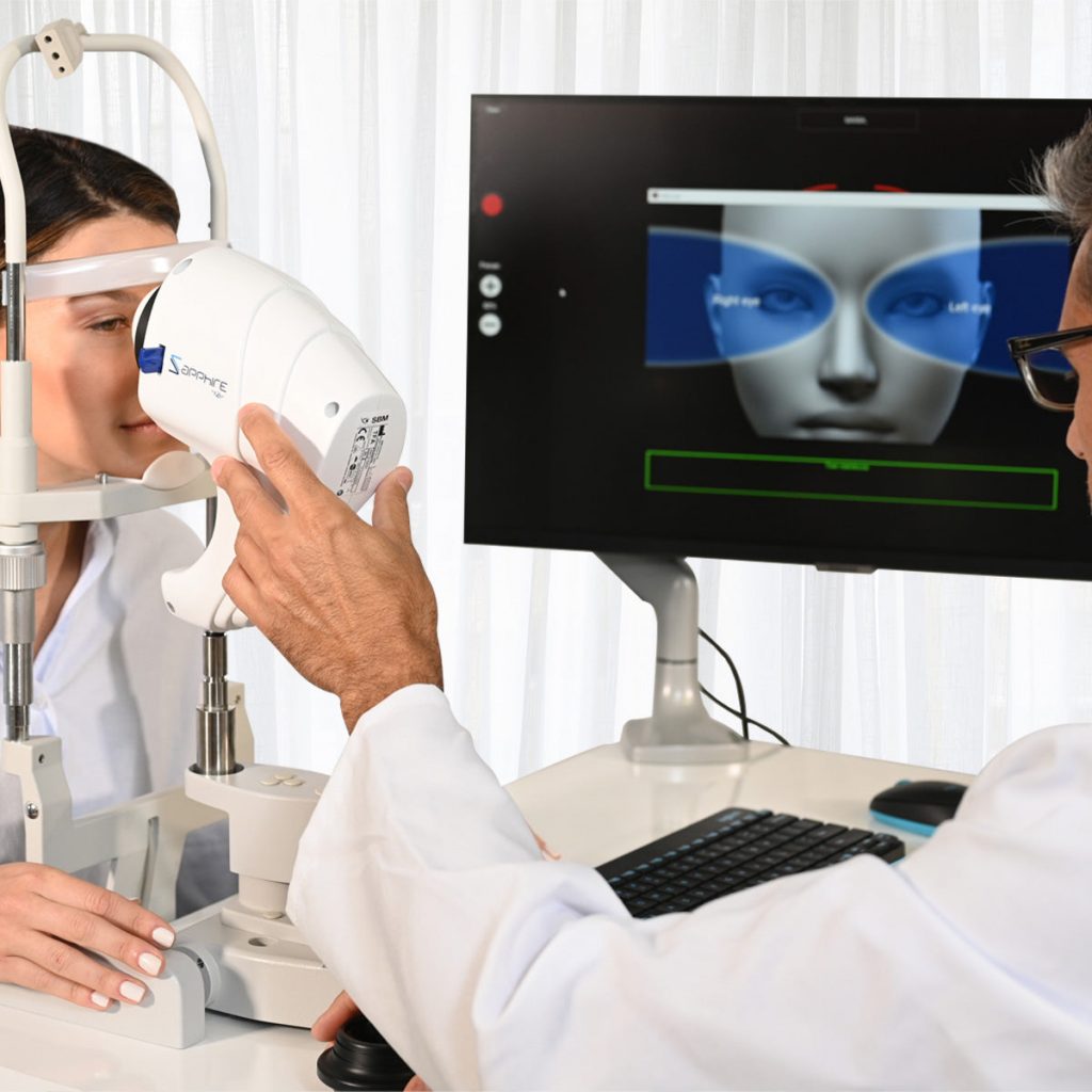
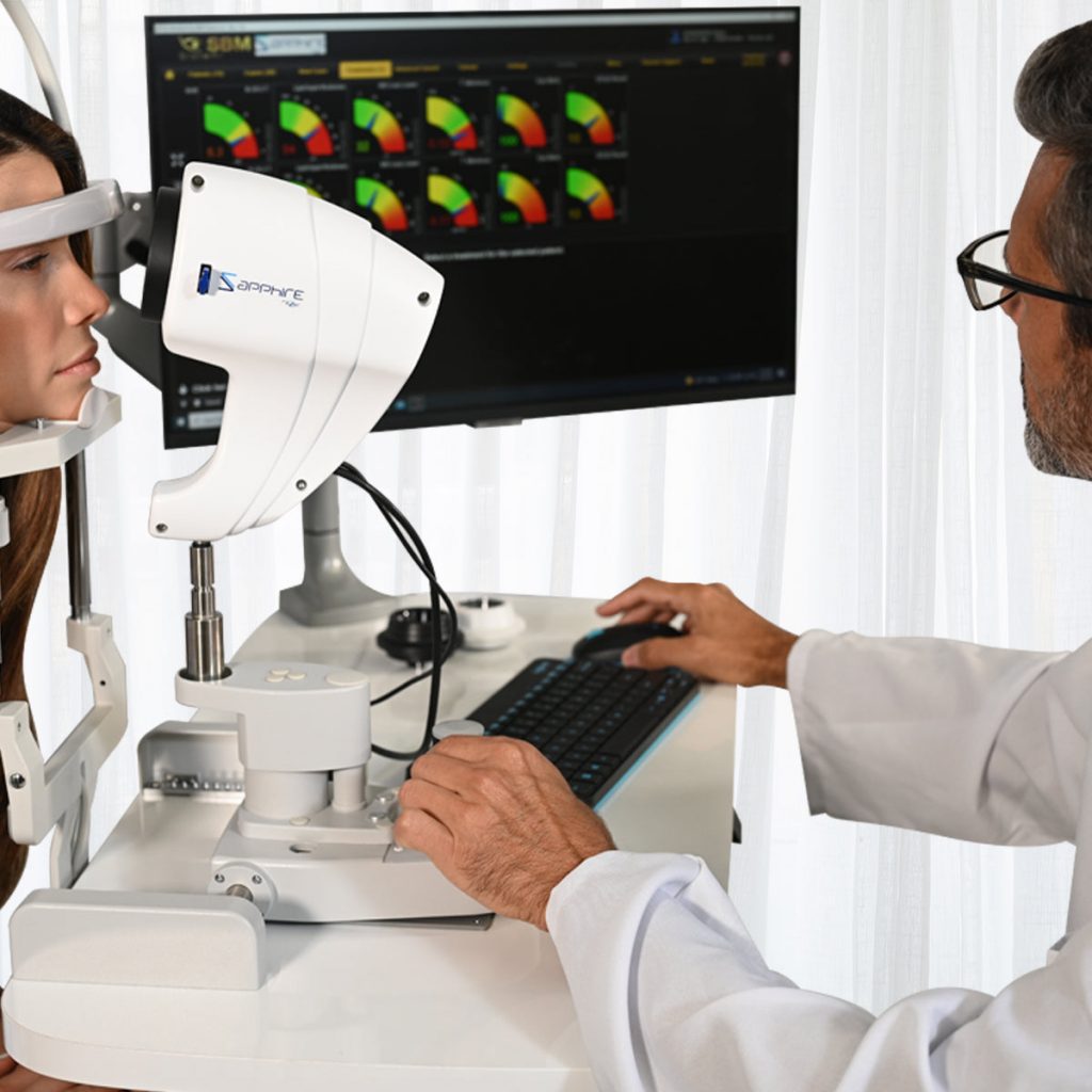
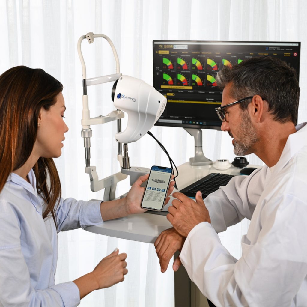
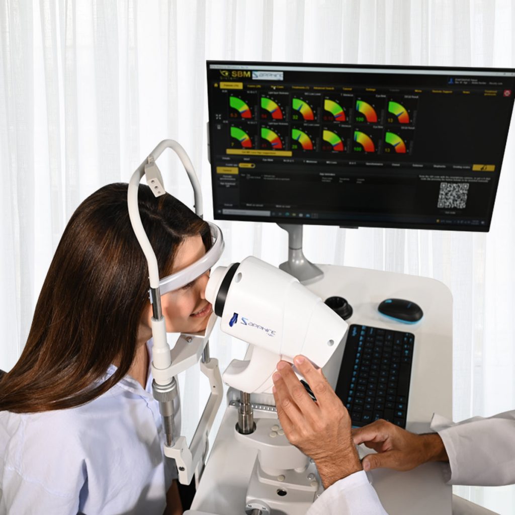
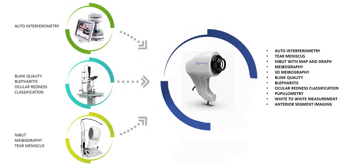
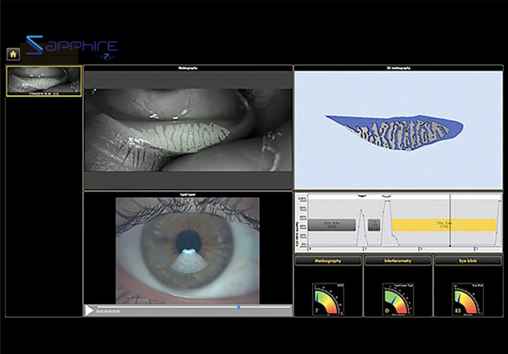
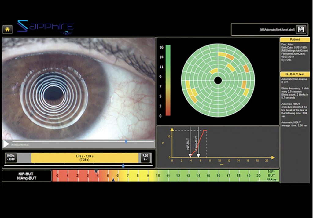
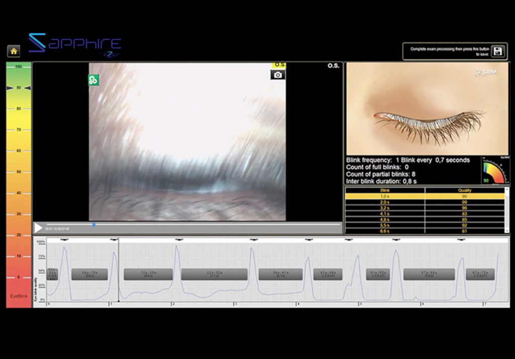
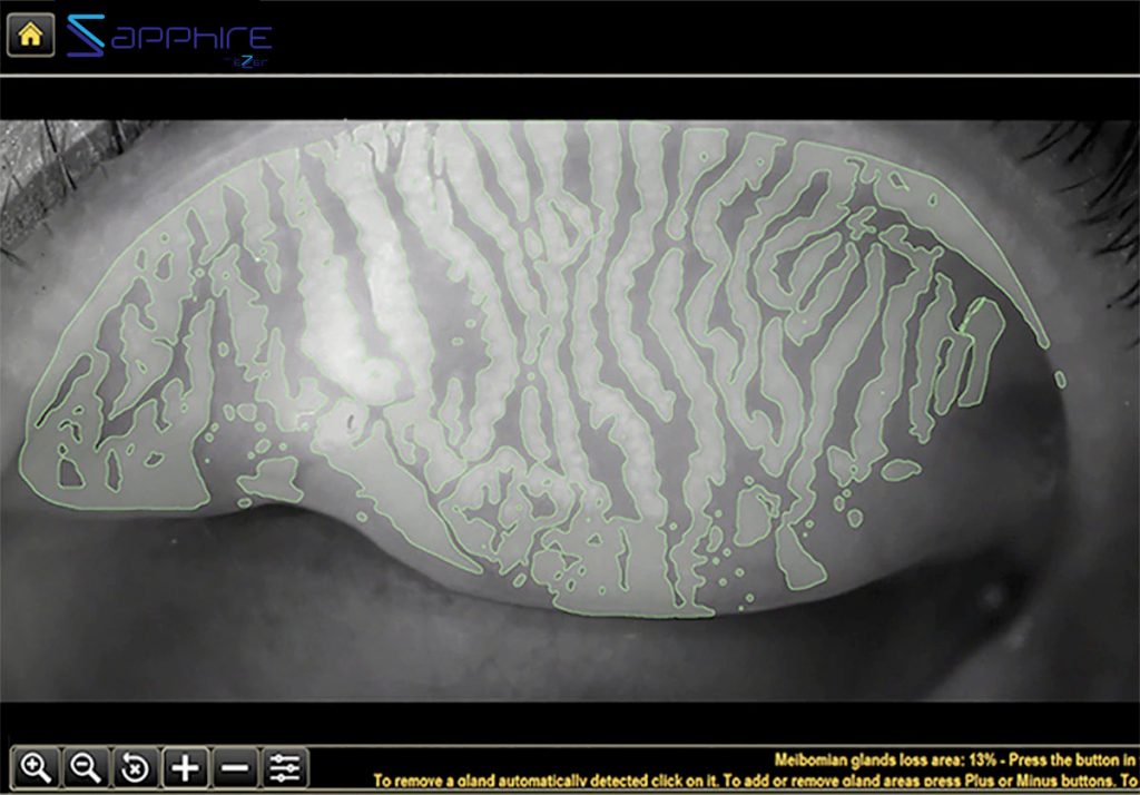
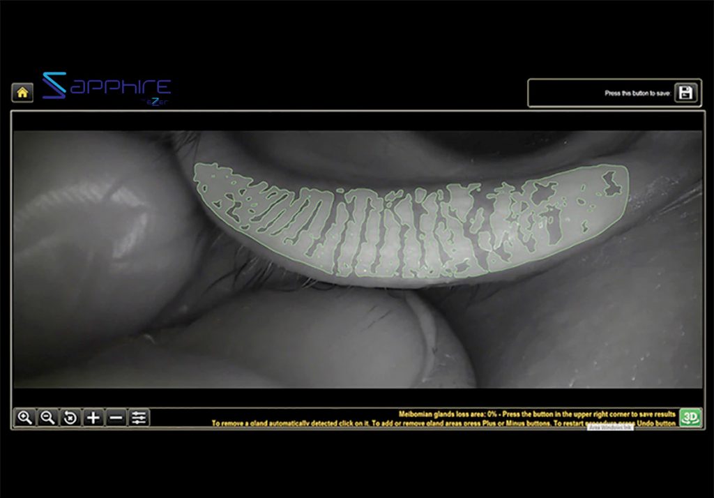
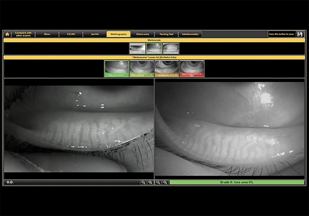
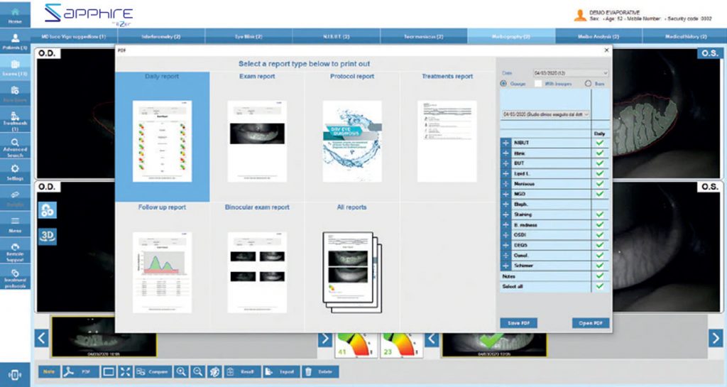
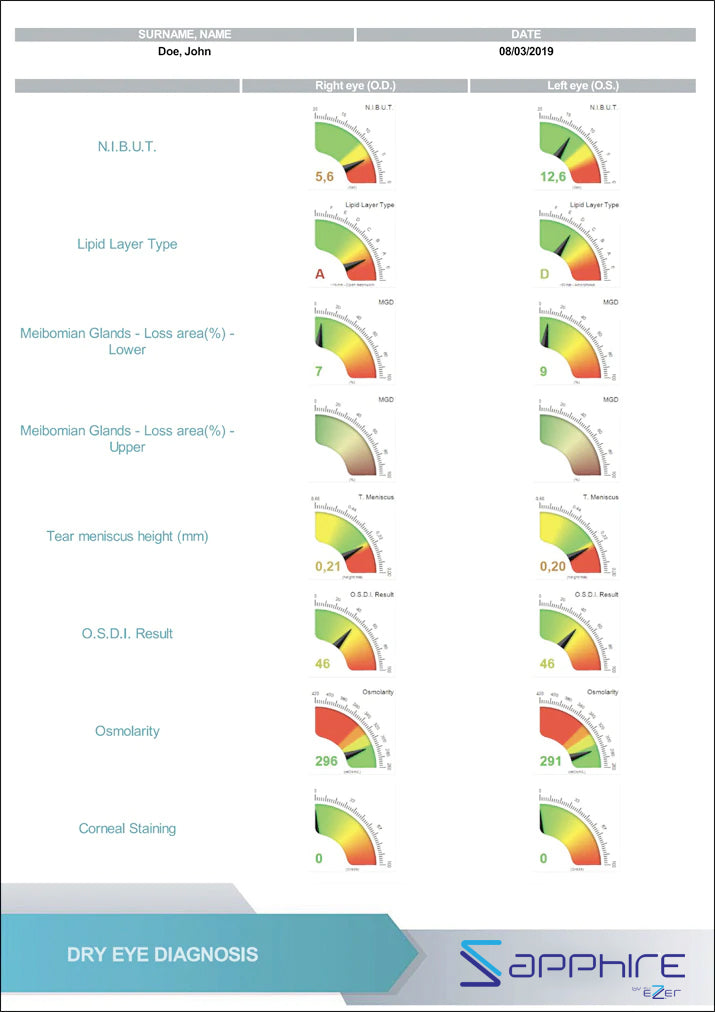
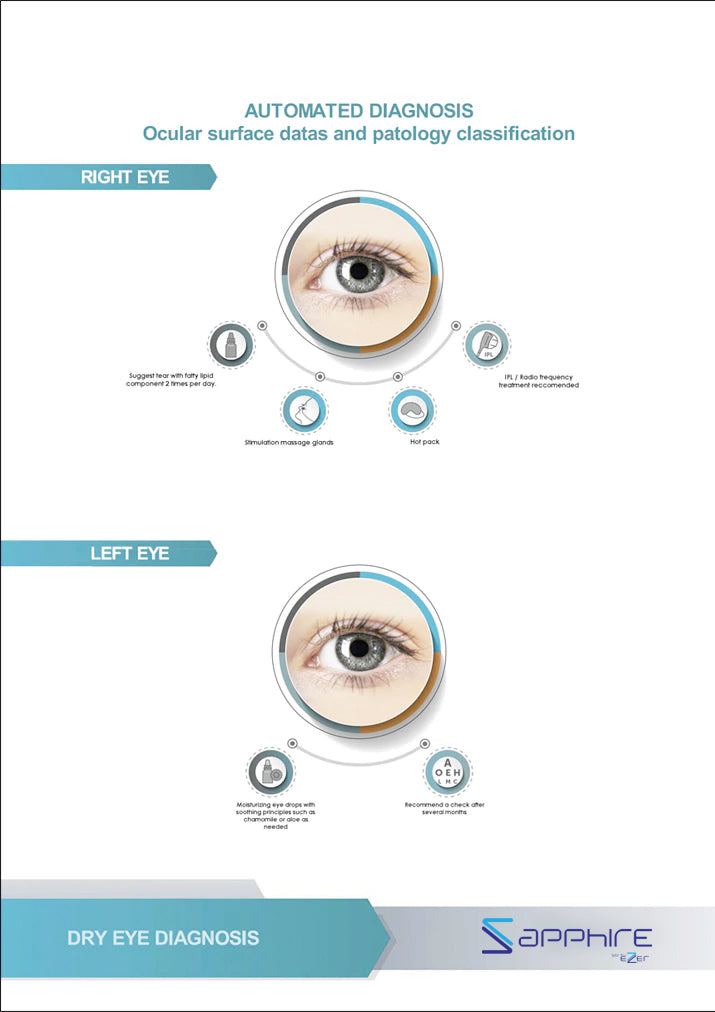
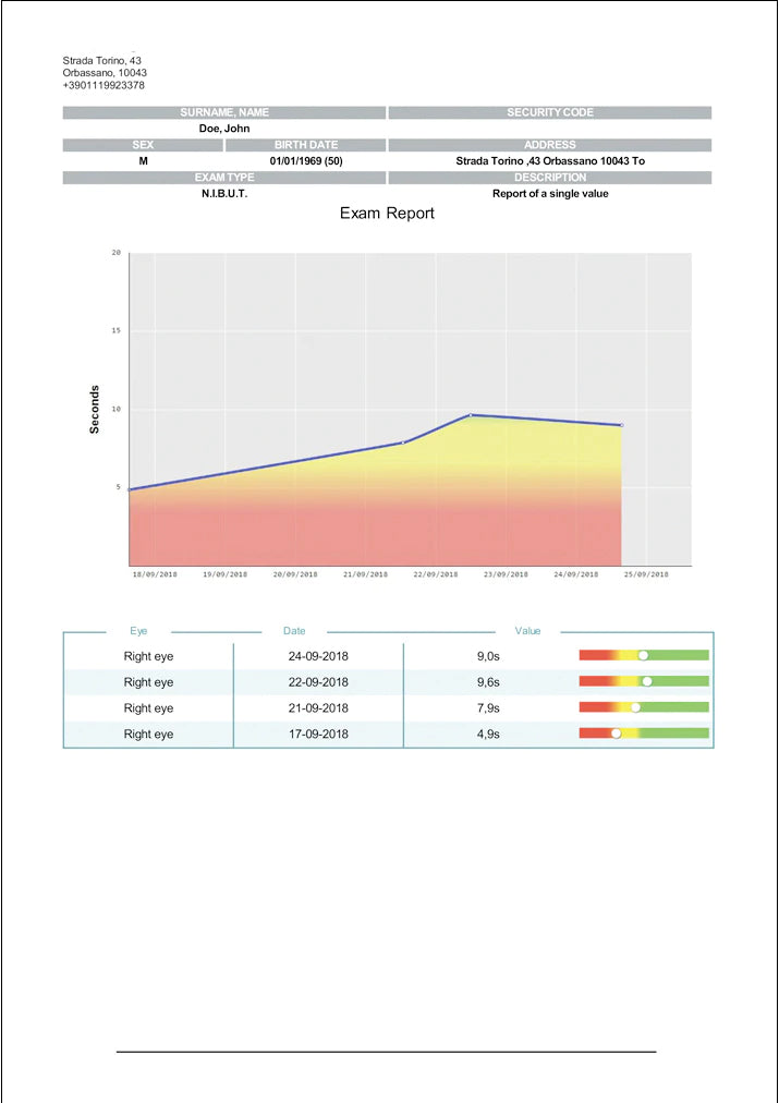
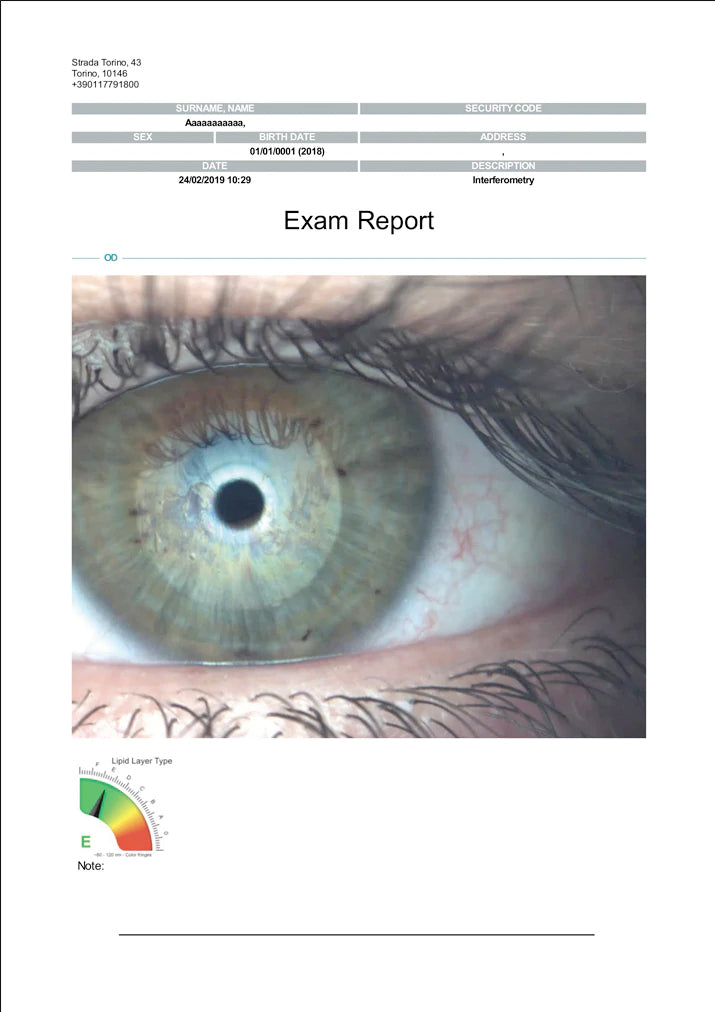
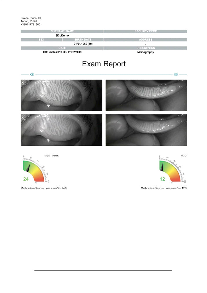
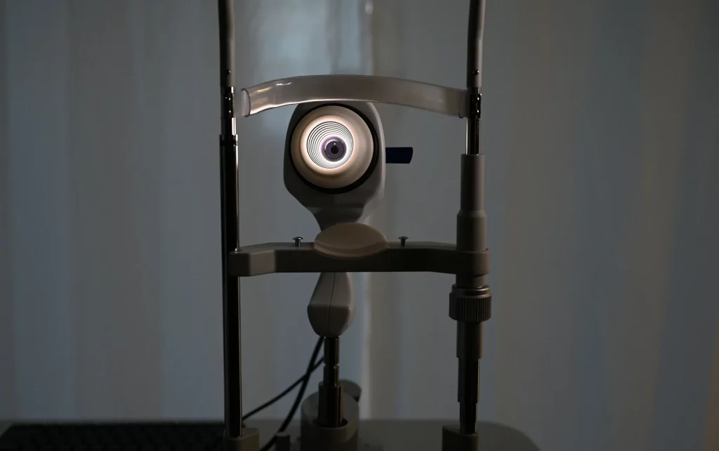
THE SAPPHIRE A+ DEVICE CAN DETECT THE LENGTH AND WIDTH OF MEIBOMIAN GLANDS VISUALIZED BY INFRARED MEIBOGRAPHY WITHOUT REQUIRING ANY INTERVENTION FROM THE USER. THE IMAGES ARE THEN CLASSIFIED AUTOMATICALLY.