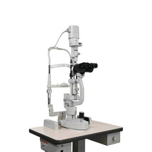
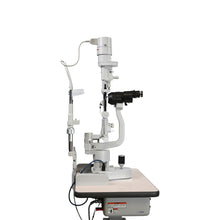
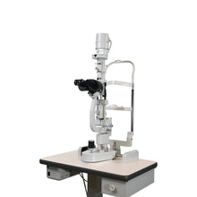
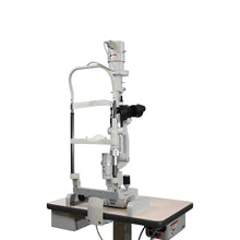
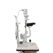
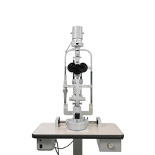
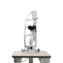
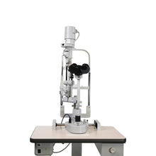
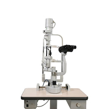
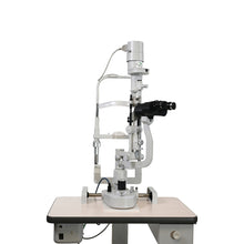
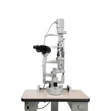
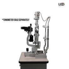
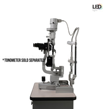
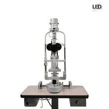
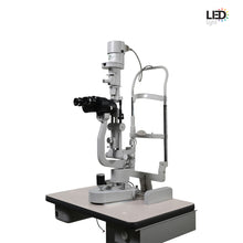
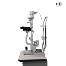
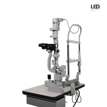
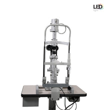
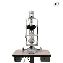
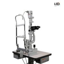
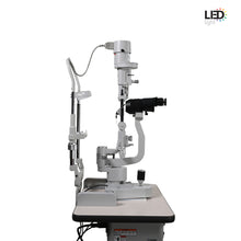
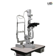
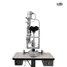
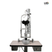
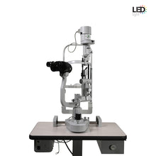
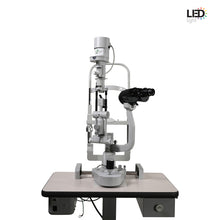
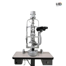
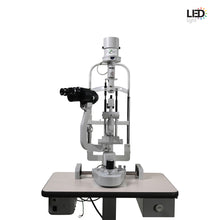
Ezer Slit Lamp ESL-Emerald-1 2X
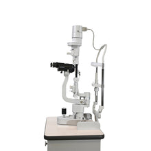
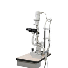
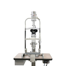
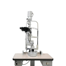
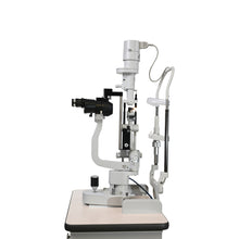
The ESL-Emerald-1 LED/Halogen “Greenough” is a first-class Japanese slit lamp instrument with an advanced Greenough Type Stereoscopic Microscope capable of delivering a precisely lit 3D view of a patient’s eye. The use of a Greenough microscope ensures that a high numerical aperture can be obtained, and that microscope maintenance is quick and simple. The basic features and user-friendly operation of the ESL-Emerald-1 LED/Halogen, along with top-quality Japanese build, make it the standard slit lamp for every practice.
| *Made in Japan |  |


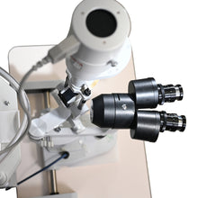
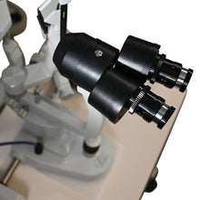
A 2-step knob provides easy manipulation of magnification power. With standard 10x and 16x magnification ratio and 10x eye piece (optional 16x), the instrument’s microscope provides quality and detail for easy detection of aberrances such as foreign objects, cysts, ulcers and allows for a clearer diagnosis of conjunctivitis, cataracts, glaucoma, macular degeneration and other diseases.

This slit lamp model offers a 13° convergent stereo view with high resolution. The pupillary distance is adjustable to a wide range (52 - 90 mm) compared to other models allowing for a more pleasant patient experience.

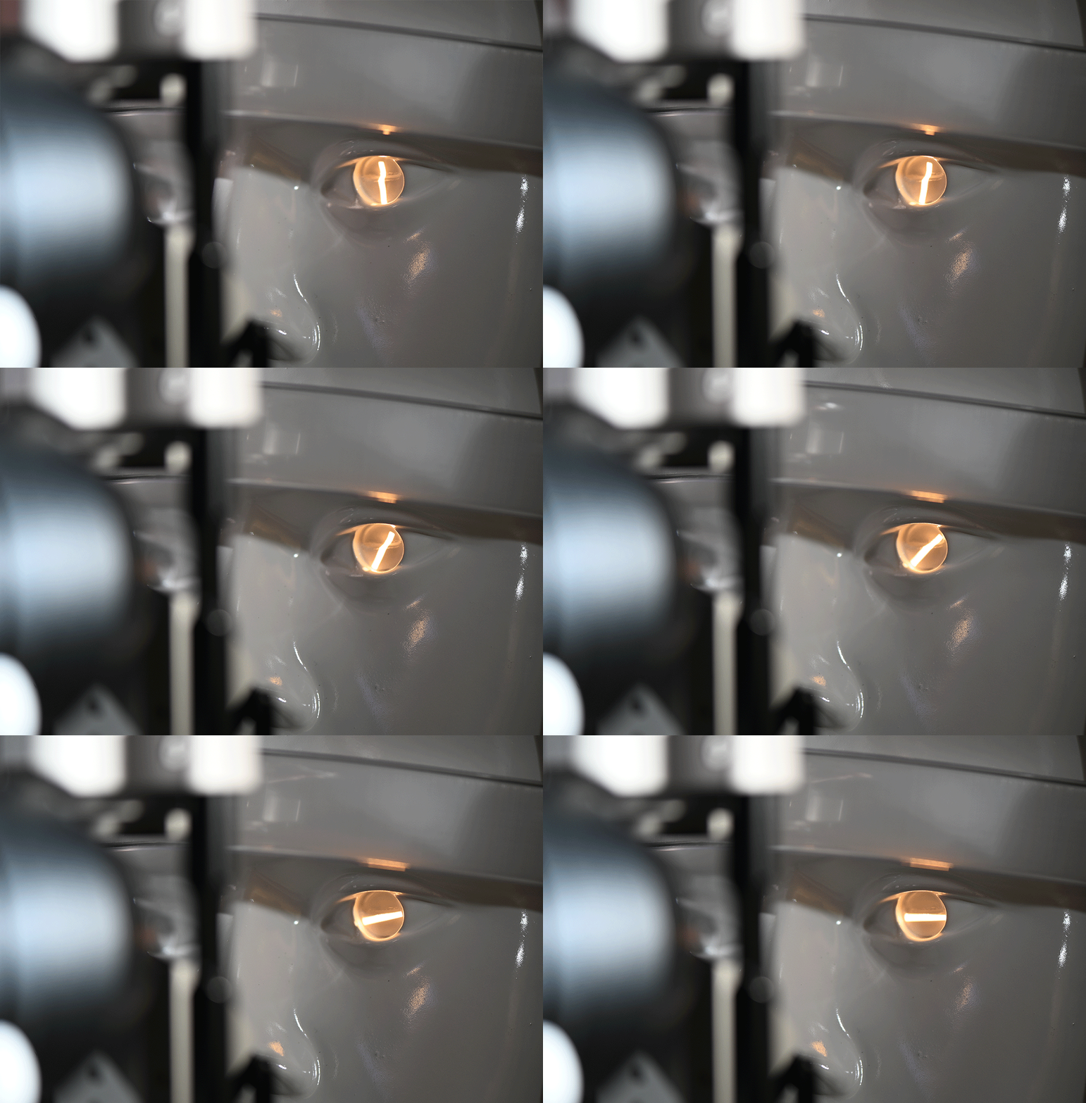
The instrument can house either a halogen or an LED lamp for tower slit illumination. Incorporating the latest LED Technology to ensure the best spectrum of light for slit lamp observation to identify eye diseases at early stages. The high illumination LED is built for longevity and decreases the energy consumption of the instrument making it the most dependable choice for daily use over many years.


With standard filters such as cobalt blue, heat absorption, red free and ½ ND, the ESL-Emerald-1 contains all the requirements for observing dry eye and corneal scarring, and the enhanced viewing of blood vessels and hemorrhages. A yellow filter (Optional) grants finer fluorescein images for detection of abnormal tear production, corneal injuries and exogenous particles. A variety of filter options are also available for upgrades.


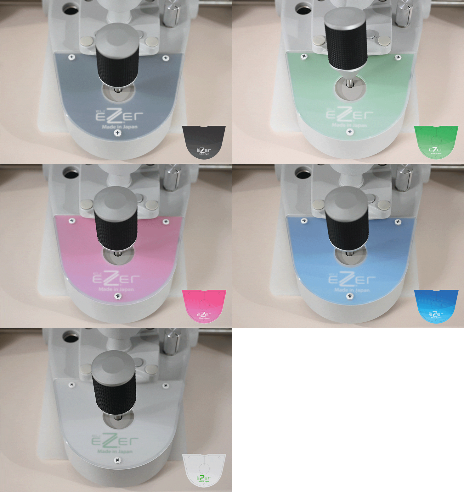
The ergonomic design allows for easy light volume control as the illumination control knob is at the base of the device and in close proximity to the joystick. The visual health doctors can make light intensity adjustments without moving their hand away from the joystick.
The base of the device houses the joystick, and its color is selectable from a variety of cover options to match your theme.

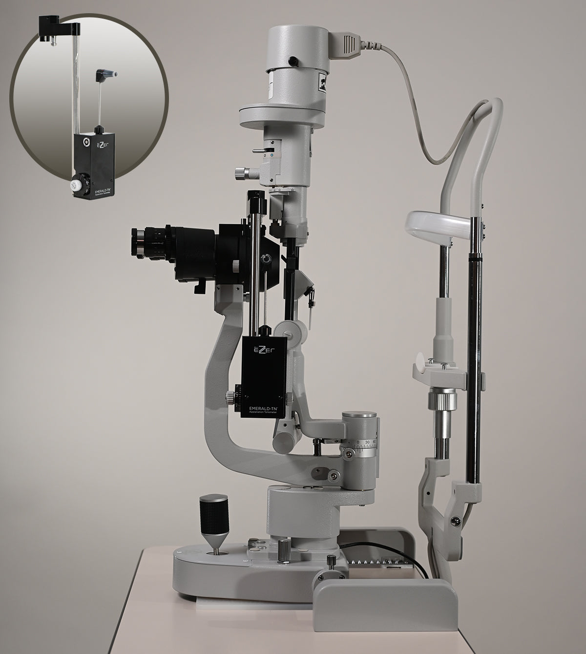
The Japanese ESL-Emerald-TN tonometer(sold separately) is a precision application tonometer designed to mount onto slit lamp equipped stereomicroscopes used for measuring intraocular pressure (IOP). The ESL-Emerald-TN works by contacting the applanation measuring cone to the eye and slowly applying pressure to the corneal surface using the measurement until it temporarily flattens.

Click here to see the manual

Click here to see the manual
| Microscope | |
| Type | Greenough Type Stereoscopic Microscope |
| Magnification Changer | 2 steps by changing knob |
| Stereo Angle | 13° |
| Eyepiece | Standard: 10x Option: 16x |
| Magnification Ratio (Field of View) | 10x (20.13mm), 16x (16.13mm) |
| Pupilary Adjustment | 52 - 90 mm |
| Focus Distance | 100 mm |
| Diopter Adjustment | +8 to -8 diopters |
| Slit Illumination | |
| Slit Width | 0 - 10mm |
| Aperture Diameter | 0.2, 1, 3, 4, 6, 10 mm |
| Slit Length | 0 - 10 mm |
| Slit Rotation | ± 90° |
| Filters |
Standard: Cobalt Blue, Heat Absorption, Red-free, 1/2 ND Option: UV, IR, 1/10 ND (to be replaced with standard filters) |
| Slit Inclination | 5° 10° 15° 20° |
| Light Source |
LED: (Orange) 14VDC 1A Halogen: Lamp bulb (12V/50W) |
| Max. Intensity (Lux) | 45,000Lux |
| Base | |
| Vertical Movement | 30 mm |
| Longitudinal Movement | 79 mm |
| Lateral Movement | 115 mm |
| Fine Base Movement | ±10 mm |
| Colour Selectable Base | Selectable |
| Chinrest | |
| Vertical Movement | 75 mm |
| Fixation Target | LED: LED 3V 0.01A (Option) Halogen: Halogen 6.3V 0.14A |
| Input Voltage | AC100, 120V, 220V, 240V 50/60 Hz |
| Power Consumption | LED: 25VA Halogen: 65VA |
| Size and Weight (without table top) | |
| Weight | 40 lbs (18.5 Kgs) |
| Dimension |
LED: 22 in(W) X 16 in(D) X 26 in(H) Halogen: 22 in(W) X 16 in(D) X 28 in(H) |


