


Ezer Ultrasound Biomicroscope EUS-2600 UBM
Go beyond the limits of Standard Ophthalmic Ultrasound Imaging with the EUS-2600 Ultrasound Biomicroscope—without sacrificing convenience or comfort!
Ultrasound became a standard diagnostic modality in almost all medical specialties including Ophthalmology. For this reason Ezer engineered an ophthalmic ultrasound imaging system with amazing charcteristics – it’s sophisticated yet easy to use, portable, and has an affordable price. The EUS-2600 UBM from Ezer combines the precision, versatility, and image quality of the EUS-2600 with cutting-edge, high-resolution biomicroscopic ultrasound technology. Moreover, its portability and convenient operating software let clinicians focus on what’s most important — delivering excellent patient care.
B-Scan & UBM
Enhanced Visibility and User-friendliness
Ophthalmic ultrasound biomicroscopy (UBM), in particular, has proved invaluable for applications ranging from diagnosing uveitis to planning phakic refractive implant surgery.
UBM utilizes 50 MHz wavelengths instead of 10 MHz—the frequency used in standard ophthalmic ultrasonography. With this advanced, non-invasive technique, clinicians and technicians can visualize the anterior ocular segment with a resolution that approaches light microscopy!
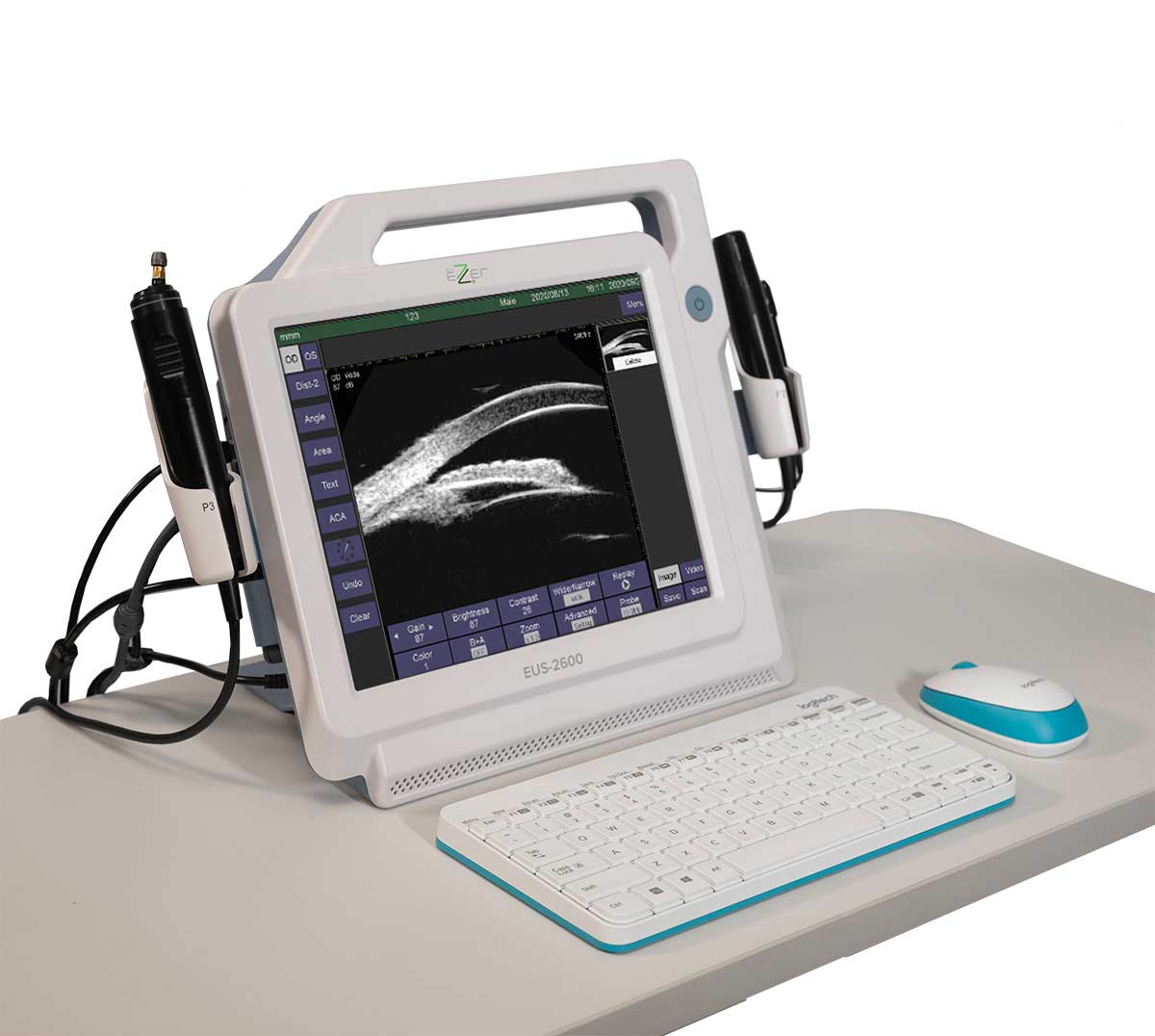
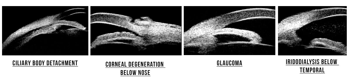
The EUS-2600 UBM is equipped with a high-resolution 50 MHz ultrasound transducer that produces two-dimensional images of the anterior segment of the eye with unparalleled detail. Examining the cornea, ciliary body, and anterior chamber angle has never been easier.
In addition, it comes with two probes for conventional ophthalmic ultrasound imaging, a 10 MHz A-scan probe and a 10 MHz B-scan probe. An optional 20 MHz high-resolution probe is available for imaging orbital fat and differentiating lesions.
The equipment captures images in multiple image / vídeo buffer slots for fast renderization alowing immediate click-and-check. The images and videos captured during the exam are saved in standard formats such as bmp and avi making them compatible with all EMR softwares.
The Ezer EUS-2600 UBM features zooming and scan depth adjustment for better observation of ocular structures and diseases. It operates in vitreous-enhanced mode for improved visibility at vitreous body.

TGC
Built-in TGC controls let users account for tissue attenuation to minimize artifacts in B-mode imaging.

A-Scan & IOL Calculation
Enhancements and Tight Integration
The EUS-2600 UBM has biometry internal algorithms for improved accuracy and reliability with averaging and standard deviation referenced against up to 10 scans per exam.
The A Scan of EUS-2600 UBM is commonly used to calculate the power of intraocular lens (IOL) implants required for cataract and refractive surgery. It can measure the axial length and makes the calculation of the intraocular lens with high precision and safety. All formulas that integrate EUS-2600 UBM have proven clinically efficient and have gained international recognition.
Image processing software
The precision probes included with the EUS-2600 UBM are driven by our advanced image processing software, which provides high-detail real-time imaging and video recording. It has six popular formulas to calculate IOL and five post-refractive IOL formulas to meet the unique conditions of individual patients.
The ultrasound has 6 popular IOL formulas and 5 post-refractive IOL formulas.
Popular IOL formulas
▪ SRK-II
▪ SRK-T
▪ BINK-II
▪ HOLLADAY
▪ HOFFER-Q
▪ HAIGIS
Post Refractive formulas
▪ History-derived
▪ Double K/SRK-T
▪ Refraction-derived
▪ ROSA
▪ SHAMMAS

Reports
The reports integrating B-scan & UBM images, A-scan/lOL results and doctor's comments with customized dictionary of symptom entries. Reports are delivered in PDF format to make printing and sharing easy. The massive storage capacity allows users to save the results of 20,000+ exams on the device, which can easily be accessed on the fly.
The compact and portable tablet design of Ezer’s EUS-2600 UBM lets clinicians bring advanced scanning technology to the exam room, saving time and space. Its built-in 12.1” high-resolution LCD touchscreen displays crisp real-time images and videos. The device’s built-in software takes advantage of the touchscreen display when inputting data in the exam room. However, a wireless mouse/keyboard combo is provided for convenient in-depth data entry. The EUS-2600 UBM also comes packaged with a foot pedal for extra convenience.
With Ezer’s commitment to imaging excellence, the EUS-2600 UBM is the ultimate solution to maximize user convenience while providing impeccable patient care.
| B-Scan & UBM | |
| Ultrasound Probes | 10MHz B-Scan Probe 50MHz UBM Probe |
| Axial Resolution |
10MHz B-Scan: ≤ 0.004in / 0.1mm UBM: ≤ 0.05in / 0.002mm |
| Lateral Resolution |
10MHz B-Scan: ≤ 0.008in / 0.2mm UBM: ≤ 0.05in / 0.002mm |
| Scan Depth |
10MHz: 1.10in - 2.36in (28mm - 60mm) 6-step Selectable |
| View Port | UBM (0.315in x 0.22in, 0.62in x 0.45in) (8mm x 5.7mm, 16mm x 11.5mm) |
| Cineloop | 10s/100 frames with dynamic replay |
| Image Acquisition | B-Scan & UBM images Snapped & Saved in real time without any limitation |
| Gray Scale | 256 Levels |
| Gain | B-Scan: 1-105dB adjustable UBM: 1-99dB |
| TGC (for B-Scan) | Default Vitreous-Enhanced Mode Customized |
| Color Codes | 8 |
| Measurements Annotation Magnification |
Distance, Area & Angle |
| Biometric A-Scan | |
| Probe | 10MHz with Fixation Red Light |
| Gain | 1-60dB |
| Measuring Method | Contact or immersion |
| Measuring Range | AL Range: 0.6in-1.58in (15mm-40mm) |
| Measuring Accuracy | ±0.002in (0.05mm) |
| Measuring Mode | Automatic (Normal, Aphakic, Special and Cataract) or Manual |
| Measurements | Average and Standard Deviation for up to 10 scans per exam Configurable Tissue Velocities under Special or Manual Mode |
| IOL Calculation | |
| General | SRK-II SRK-T BINK-II HOLLADAY HOFFER-Q HAIGIS |
| Post Refractive | History-derived Double K/SRK-T Refraction-derived ROSA SHAMMAS |
| General | |
| Display | High Resolution 12.1"" LCD |
| Printer Compatibility | Graph/Text Printer and Video Printer (PAL) |
| Interface | Video-Out (PAL) HDMI USB 2.0 Ports |
| Operations | Touch Screen Wireless Mouse & Keyboard Footswitch |
| HDD | 500GB or higher |
| Network | Folder/Report Sharing |
| Power Supply | AC-100-240V, 50/60Hz |
| Optional | Eye cup Immersion Shell Video Thermal Print |
| B-Scan & UBM | |
| Ultrasound Probes | 10MHz B-Scan Probe 50MHz UBM Probe |
| Axial Resolution |
10MHz B-Scan: ≤ 0.004in / 0.1mm UBM: ≤ 0.05in / 0.002mm |
| Lateral Resolution |
10MHz B-Scan: ≤ 0.008in / 0.2mm UBM: ≤ 0.05in / 0.002mm |
| Scan Depth |
10MHz: 1.10in - 2.36in (28mm - 60mm) 6-step Selectable |
| View Port | UBM (0.315in x 0.22in, 0.62in x 0.45in) (8mm x 5.7mm, 16mm x 11.5mm) |
| Cineloop | 10s/100 frames with dynamic replay |
| Image Acquisition | B-Scan & UBM images Snapped & Saved in real time without any limitation |
| Gray Scale | 256 Levels |
| Gain | B-Scan: 1-105dB adjustable UBM: 1-99dB |
| TGC (for B-Scan) | Default Vitreous-Enhanced Mode Customized |
| Color Codes | 8 |
| Measurements Annotation Magnification |
Distance, Area & Angle |
| Biometric A-Scan | |
| Probe | 10MHz with Fixation Red Light |
| Gain | 1-60dB |
| Measuring Method | Contact or immersion |
| Measuring Range | AL Range: 0.6in-1.58in (15mm-40mm) |
| Measuring Accuracy | ±0.002in (0.05mm) |
| Measuring Mode | Automatic (Normal, Aphakic, Special and Cataract) or Manual |
| Measurements | Average and Standard Deviation for up to 10 scans per exam Configurable Tissue Velocities under Special or Manual Mode |
| IOL Calculation | |
| General | SRK-II SRK-T BINK-II HOLLADAY HOFFER-Q HAIGIS |
| Post Refractive | History-derived Double K/SRK-T Refraction-derived ROSA SHAMMAS |
| General | |
| Display | High Resolution 12.1" LCD |
| Printer Compatibility | Graph/Text Printer and Video Printer (PAL) |
| Interface | Video-Out (PAL) HDMI USB 2.0 Ports |
| Operations | Touch Screen Wireless Mouse & Keyboard Footswitch |
| HDD | 500GB or higher |
| Network | Folder/Report Sharing |
| Power Supply | AC-100-240V, 50/60Hz |
| Optional | Eye cup Immersion Shell Video Thermal Print |

